
How Is a Digital Mammogram Done?
What Patients Should Know Before a Digital Mammogram
A Digital Mammogram is a breast imaging examination that uses X-ray technology and digital detectors to create detailed images of breast tissue. Many patients know that mammograms are used for breast cancer screening, but they may still have questions about what happens during the examination.
The digital mammogram procedure is usually completed within a short time. A trained technician guides the patient through each step, from preparation to image capture. Unlike traditional film mammography, Digital Mammography stores images electronically, which allows radiologists to review and analyze them more efficiently.
Knowing how a Digital Mammogram is performed can help patients feel more prepared and understand what to expect during the appointment.
Preparation Before the Examination
Before a Digital Mammogram, patients usually receive a few simple instructions.
On the day of the examination, it is recommended to avoid using deodorant, powder, lotion, or other products around the breasts and underarm areas. Some ingredients in these products may appear as small spots on the images and could affect image interpretation.
Patients should also wear comfortable clothing because they will need to remove clothing from the upper body before imaging. A two-piece outfit is often more convenient for the examination.
Before the procedure begins, the technician may ask about previous breast examinations, breast surgery, symptoms, or other relevant medical information. Patients who are pregnant or may be pregnant should inform the healthcare provider before the examination.
These preparation steps help ensure that the images are clear and that the healthcare team has the information needed for accurate evaluation.
During the Digital Mammogram Procedure
During the examination, the patient stands in front of the digital mammography system. The technician carefully positions one breast on the detector platform and adjusts the equipment to capture the required areas.
The breast is then gently compressed between two plates. This step is an important part of the digital mammography process because it helps spread breast tissue and reduce overlapping structures.
Although compression may feel uncomfortable, it usually lasts only a few seconds. It helps reduce movement during imaging, improve image clarity, and allow the system to obtain high-quality images with an appropriate radiation dose.
The technician may reposition the patient several times to capture images from different angles. These views provide a more complete evaluation of breast tissue.
Breast Compression and Image Capture
After proper positioning, the system uses a small amount of X-ray radiation to capture images of the breast.
The digital detector collects the X-ray information and converts it into electronic signals. The system processes these signals to create digital breast images that can be viewed on a workstation.
Compared with traditional film mammography, Digital Mammography allows faster image access and easier image management. Radiologists can adjust image display settings, enlarge specific areas, and compare current images with previous examinations when available.
The quality of the final images depends on several factors, including patient positioning, breast compression, equipment performance, and the experience of the healthcare team.
Image Review and Follow-up After the Examination
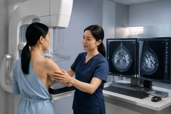
After the images are captured, the technician checks whether they meet quality requirements. The images need to show enough breast tissue and have appropriate exposure levels before they are sent for review.
A radiologist then evaluates the images for possible abnormalities. The review process may include looking for changes in breast tissue, suspicious areas, or findings that require further evaluation.
In some cases, additional imaging may be recommended. This does not always mean that cancer is present. Additional views, ultrasound, or other examinations may simply provide more information about an area that needs closer evaluation.
After the Digital Mammogram, most patients can return to their normal activities immediately. The healthcare facility will provide the results according to its reporting process.
Conclusion: Understanding the Digital Mammography Process
The digital mammogram procedure is a simple and efficient breast imaging process that combines X-ray technology, digital detectors, and professional interpretation.
From preparation and positioning to image capture and review, each step helps create clear images for breast health evaluation. Understanding the process can help patients feel more comfortable and prepared before their examination.
Digital Mammography continues to play an important role in modern breast cancer screening. Need a new Digital Mammography system? Contact us for professional solutions and technical support.

