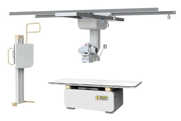
How Does Digital Radiography Work?
Digital Radiography creates digital medical images by combining X-ray technology with digital detectors and computer-based image processing.
The imaging process involves several stages, including X-ray transmission, digital detection, signal conversion, image processing, and image management. Together, these technologies enable healthcare providers to capture, view, and manage medical images within modern digital imaging environments.
Understanding how Digital Radiography works helps healthcare providers evaluate imaging technologies. It also helps them better understand how modern digital imaging systems support clinical and operational workflows.
The Basic Principle of Digital Radiography
Digital Radiography uses X-rays to capture information from inside the body. During an examination, X-rays pass through the patient and interact with different anatomical structures.
Different tissues attenuate X-ray beams at different rates. Dense structures such as bones absorb more X-ray energy, while soft tissues allow more X-rays to pass through. These differences create the image contrast required for medical image formation.
The transmitted X-rays contain anatomical information. This information can be captured and converted into a digital image through the Digital Radiography system.
The Digital Radiography Workflow
Understanding how Digital Radiography works begins with examining the key stages involved in image acquisition and processing.
X-ray Generation and Transmission
The imaging process starts with the X-ray generator, which produces a controlled X-ray beam and directs it toward the area being examined.
During exposure, X-rays pass through the patient’s body and interact with different tissues and anatomical structures. Because different tissues have different densities and compositions, they absorb X-ray energy at different levels.
For example, dense structures such as bones absorb more X-ray energy, while soft tissues such as muscles and fat allow more X-rays to pass through. These differences in attenuation create the contrast information required for image formation. They also provide the foundation for visualizing internal anatomical structures.
After passing through the body, the transmitted X-rays carry anatomical information. They then continue toward the digital detector, where image acquisition begins.
Digital Detection and Signal Conversion
Once the transmitted X-rays reach the detector, they are captured and converted into usable imaging data. The digital detector is one of the most important components of a Digital Radiography system because it transforms X-ray energy into electronic signals.
As X-rays strike the detector surface, the detector records the radiation intensity received at different locations. Because different tissues absorb X-rays differently, the radiation reaching each area also varies.
The detector converts these differences into electronic signals and then into digital data. Each data point corresponds to a specific location within the examined anatomy. This creates the information required for image reconstruction.
The acquired digital data is then transferred to the computer system for processing and analysis. Because detector performance directly influences image quality, digital detectors have become a core component of modern Digital Radiography systems.
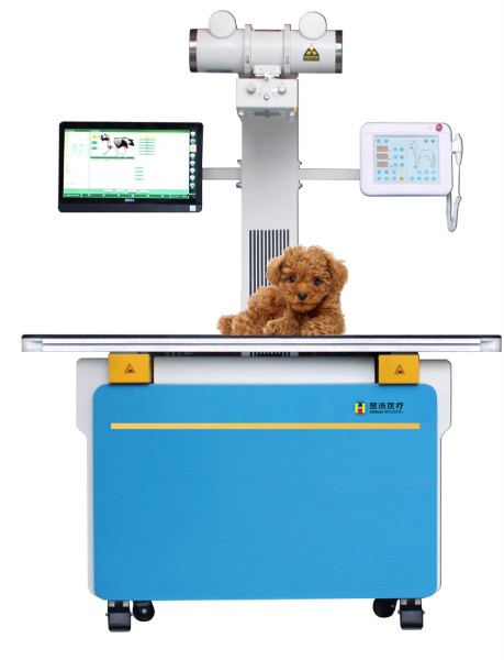
Image Processing and Display
After image data has been acquired, image processing software analyzes and reconstructs the information to generate a digital medical image.
During this stage, the system reconstructs image content based on detector data. It converts raw signals into a visual representation of the examined anatomy. Through digital processing, raw data becomes easier to view, interpret, and manage.
Modern Digital Radiography systems use image processing technologies to optimize image presentation. They also support clinical review and improve imaging workflow efficiency. Advanced algorithms enhance image contrast and improve anatomical detail visibility.
This helps healthcare providers evaluate images more effectively.
The reconstructed image is displayed on a workstation.Healthcare professionals can view, enlarge, measure, and analyze the image. Because the images are digital, they can be transferred efficiently between systems. This helps healthcare facilities streamline imaging management processes.
Digital Image Storage and Management
After a digital image has been generated, it can be stored and managed in digital form.
Unlike film-based imaging, Digital Radiography stores image data digitally. This makes storage, retrieval, and management more efficient.
Healthcare facilities can quickly access imaging records when needed and support more streamlined clinical workflows.
Digital image management supports storage, retrieval, and sharing of imaging records.This improves overall workflow efficiency.
Conclusion
Digital Radiography works by combining X-ray imaging with digital detector technology and computer-based image processing to create medical images that support clinical evaluation and patient care.
By enabling digital image acquisition, processing, storage, and management, Digital Radiography has become an important part of modern healthcare imaging environments across hospitals, clinics, imaging centers, and Veterinary Digital Radiography applications. These capabilities help healthcare facilities improve workflow efficiency and support the ongoing transition to digital imaging.
As digital imaging technology continues to advance, Digital Radiography remains an important part of modern healthcare services, helping providers improve imaging efficiency and workflow management.
SEEHO offers Digital Radiography solutions designed to support evolving clinical needs.