
Digital Mammogram Maintenance Checklist: How to Prevent Downtime and Extend Equipment Life
Preventing unscheduled downtime and prolonging the working life of a clinical digital mammography system comes down to three daily and weekly habits: executing morning flat-field calibrations, verifying mechanical paddle torque, and tracking tube heat dissipation. Neglecting these basic checks leads directly to ghosting artifacts, mechanical motor jams, and premature tube replacement. Establishing a systematic preventive maintenance protocol protects your imaging hardware, satisfies statutory inspection guidelines, and keeps screening rooms running on schedule.
Daily Quality Control Protocols for Your Digital Mammography System
Technologists must avoid taking patient exposures immediately after boot-up. Digital flat-panel detectors require sufficient warm-up time to reach stable operating temperatures. Skipping this preparation on a digital mammography system produces baseline signal drift that obscures subtle tissue contrast.
Phantom Image Testing and Artifact Evaluation
Every morning shift must start with an accredited mammography phantom exposure:
- Object Visibility: Check baseline scoring sheets for fibers, microcalcification specks, and low-contrast tumor-like masses. If visibility drops below required QA thresholds, pause clinical exams for recalibration.
- Flat-Field Inspection: Examine uniform exposures with narrow display window settings to detect dead pixel columns, light-guide dust, or fine scratches on compression plates.
- Quadrant Balance: Verify signal value consistency across all four image quadrants to confirm uniform panel response.
Detector Flat-Field Calibration and System Warm-Up
A cold detector matrix creates image ghosting across back-to-back exposures. Technologists should initiate the automated tube warm-up program to distribute thermal loads evenly across the anode track. Once the focal track warms up, running a software-guided flat-field calibration resets dark current offset values and equalizes pixel gain across the panel.
Monthly and Semi-Annual Mechanical & Electrical Checks
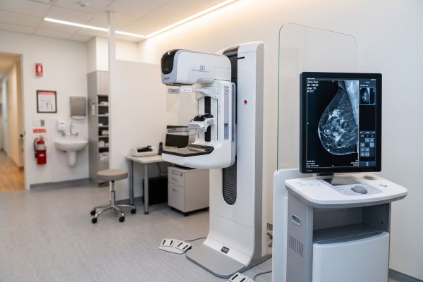
Subtle mechanical misalignments and cable fatigue typically cause patient positioning complaints long before triggering emergency error codes on a digital mammography system.
| Inspection Interval | Target Subsystem | Standard Maintenance Action and Tolerance |
| Monthly | Compression Mechanism | Measure automatic cutoff force (Standard: 150 N – 200 N / 33–45 lbs) |
| Monthly | Emergency Release | Test foot switches and manual override knobs for instant paddle release |
| Semi-Annual | Gantry C-Arm Assembly | Confirm rotational angle accuracy ($\pm 1^{\circ}$) and magnetic brake hold |
| Semi-Annual | High-Voltage Cabling | Check dynamic flex tracks for cable jacket wear, strain, or oil leaks |
| Semi-Annual | Collimator Light Field | Confirm light field matches radiation exposure boundaries ($<2\%$ of SID) |
Compression Force Calibration and Emergency Release Verification
Adequate breast compression flattens tissue structures and lowers radiation dosage, but incorrect motor torque compromises patient safety:
- Force Gauge Measurement: Use a dedicated digital scale to confirm motorized compression cuts off between 111 N and 200 N. If automatic drive force exceeds 200 N, remove the paddle from service.
- Emergency Release Testing: Depress the emergency stop button during active compression. The paddle must back off instantly without sticking, and the manual mechanical release knob must rotate smoothly.
Gantry Rotation, Brake Stability, and Cable Integrity
Repeated angle rotations during routine diagnostic views cause mechanical wear across internal drive belts:
- Brake Holding Force: Position the C-arm at a $45^{\circ}$ angle and apply steady hand pressure. If the electromagnetic brake slips, clean the brake disk and adjust pad clearance.
- Cable Track Inspection: Inspect internal dynamic cable carriers inside the lower column. Plastic shavings or jacket powder indicate cables are rubbing against metal housing walls.
Climate and Humidity Monitoring for FPD Protection
Direct-conversion amorphous Selenium (a-Se) detectors are sensitive to room environment fluctuations:
- Temperature Stability: Keep HVAC systems running continuously to maintain ambient room temperatures between 20°C and 25°C.
- Humidity Control: Maintain relative humidity between 30% and 60% non-condensing to prevent electronic corrosion and selenium crystallization.
Protecting High-Cost Components in a Digital Mammography System
The flat-panel detector and X-ray tube make up more than half the hardware value of any digital mammography system. Protecting these assemblies requires clean utility power and disciplined thermal management.
X-Ray Tube Thermal Management and Exposure Logging
Heavy screening schedules create high thermal stress on X-ray tube assemblies:
- Heat Unit Tracking: Keep an eye on anode heat unit percentages on the console. Pause exposures for two minutes if heat accumulation crosses 75%.
- Chiller Maintenance: Clean dust off radiator cooling fins every six months and inspect closed-loop coolant sight glasses for bubbles or discoloration.
- Cumulative mAs Records: Record exposure counts regularly to anticipate filament wear and budget for tube replacements before catastrophic vacuum failures occur.
Power Conditioning and UPS Battery Maintenance
Unfiltered line voltage damages sensitive thin-film transistor arrays. Connecting your digital mammography system to an online double-conversion UPS isolates digital readout electronics from line transients and power sags. In addition, technicians should run semi-annual battery discharge tests to verify at least 15 minutes of backup power for completing running views and executing safe software shutdowns.
Practical QA Implementation for Reliable Imaging
Catching detector drift or mechanical wear early avoids costly emergency repair dispatches. Adding these daily and monthly verification steps to your standard department opening procedures ensures reliable uptime, consistent image sharpness, and full regulatory compliance.
Contact our technical support engineers to access standardized quality assurance templates, order certified replacement components, or schedule complete preventive servicing for your digital mammography system.

