What Factors Affect Dynamic Digital Radiography Image Quality?
Dynamic digital radiography image quality is determined by an interconnected chain of technical and operational factors, including flat-panel detector specifications, radiographic exposure parameters, patient positioning stability, real-time post-processing algorithms, and overall system configuration. Rather than resulting from a single isolated hardware metric, diagnostic clarity in continuous cine sequences depends on how effectively hardware components synchronize with procedural protocols. Dynamic digital radiography captures continuous anatomical movement over time, meaning spatial sharpness and temporal motion fidelity must be balanced simultaneously. Understanding how these distinct variables interact enables imaging facilities to optimize diagnostic visualization while strictly adhering to radiation safety principles.
Detector Performance Has a Major Impact
The flat-panel detector serves as the primary component in the digital imaging chain, governing initial X-ray photon capture and signal conversion efficiency.
A detector with a refined pixel pitch provides the baseline spatial resolution necessary to delineate fine cortical bone margins and subtle lung parenchymal patterns. Concurrently, high detective quantum efficiency ensures optimal signal retention at low radiation levels, suppressing electronic background noise. Furthermore, rapid panel refresh speeds and uniform scintillator response are indispensable for dynamic digital radiography. If detector lag or residual ghosting occurs between consecutive frames, rapid physiological movement will generate severe temporal blur that degrades edge delineation.
Exposure Parameters Regulate Signal and Dose Balance
Radiographic technique settings directly influence image contrast, signal-to-noise ratio, and overall patient radiation exposure during multi-frame cine acquisitions.
- Tube Voltage (kVp): Determines beam penetration and subject contrast across dense skeletal and soft tissue structures.
- Tube Current-Time Product (mAs): Governs total photon quantity per exposure pulse, directly influencing background noise.
- Pulsed Width and Frame Rate: Controls the temporal freezing capacity needed to capture rapid anatomical excursions.
Balancing these technical settings is vital in dynamic digital radiography. Setting exposure values too low introduces excessive quantum mottle that obscures delicate tissue borders. Conversely, excessive exposure unnecessarily inflates cumulative radiation dose without conferring commensurate diagnostic benefit.
| Technical Variable | Operational Determinant | Direct Clinical & Diagnostic Impact |
| Detector Hardware | Pixel size, refresh rate, and detective quantum efficiency | Baseline structural sharpness, low-noise fidelity, and ghosting elimination |
| Exposure Control | Pulsed mAs, tube potential (kVp), and exposure duration | Penetration depth, quantum mottle suppression, and cumulative dose management |
| Kinetic Control | Immobilization, physiological gating, and breathing coaching | Motion artifact minimization and true functional biomechanical tracking |
| Algorithmic Engine | Recursive noise filtering and multi-frequency contrast enhancement | Enhanced edge visibility and visualization across high-speed dynamic frames |
Patient Motion and Positioning Alter Dynamic Digital Radiography Results
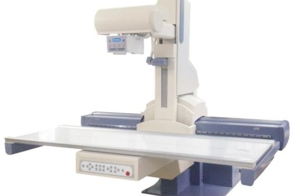
Because dynamic digital radiography evaluates active physiological kinetics, differentiating intentional diagnostic movement from uncontrolled extraneous motion is essential for image clarity.
Unintended patient sway, tremor, or inconsistent respiration can introduce severe motion artifacts that obscure structural interfaces. Proper patient positioning aids and clear breathing instructions ensure that recorded kinematics reflect true physiological behavior. In joint stress studies or thoracic motion evaluations, technologists must secure stable alignment so that recorded dynamic sequences highlight targeted biomechanical anomalies rather than erratic positioning shifts.
Image Processing Influences the Final Cine Presentation
Advanced digital image processing software converts raw detector data into high-contrast diagnostic cine sequences suitable for clinical review.
Sophisticated processing engines deploy recursive noise filtering, multi-scale contrast adjustment, and dynamic range equalization in real time. These algorithms optimize visual clarity between varying tissue densities without compromising delicate structural boundaries. However, digital post-processing cannot fully compensate for hardware limitations or poor exposure calibration. If a dynamic digital radiography unit captures raw data degraded by severe motion blur or underexposure, aggressive software sharpening will merely amplify background artifacts.
System Configuration and Examination Protocols in Dynamic Digital Radiography
The ultimate image quality delivered by dynamic digital radiography depends on systemic engineering harmony and proper clinical protocol selection.
A high-output X-ray tube with rapid heat dissipation must support sustained pulsed cine runs without triggering thermal throttling. Similarly, mechanical gantry stability prevents vibrational artifacts during motorized movements. Aligning frame acquisition rates with specific anatomical velocities—such as utilizing higher frame rates for cardiac or swallowing studies and moderate rates for diaphragmatic sweeps—ensures optimal diagnostic outcomes across varied departmental workflows.
Conclusion
Diagnostic image quality in dynamic digital radiography is governed by the coordinated interaction of flat-panel detector efficiency, precise exposure calibration, patient motion stabilization, and real-time processing software. Clinical teams cannot assess performance through a single hardware parameter alone. Hospital procurement committees and radiology departments should evaluate how effectively the entire radiographic chain integrates to deliver consistent, low-dose dynamic imaging across routine clinical workloads.
Contact our radiographic specialists today to review imaging configurations and explore dynamic digital radiography solutions for your facility.