Is Dynamic Digital Radiography Safe for Patients?
Dynamic digital radiography is safe for clinical use when conducted under valid medical indications, optimized pulsed exposure parameters, and standardized operational workflows, though the ionizing radiation inherent to X-ray imaging requires systematic management. Patient safety in dynamic imaging does not stem solely from hardware build quality; it is a shared outcome of equipment design, appropriate protocol selection, and careful technologist technique. Because dynamic digital radiography captures sequential frames rather than an isolated still exposure, radiology departments must treat patient protection as an active operational process. Balancing diagnostic visual data with rigorous exposure controls ensures that institutions maximize clinical benefit while holding radiation risks to acceptable baselines.
Understanding Radiation Exposure During Dynamic Imaging
Because dynamic dr relies on diagnostic X-rays, evaluating safety begins with how energy is delivered to patient tissues. Unlike static radiography where exposure lasts a fraction of a second, dynamic cine acquisitions deliver repeated bursts of photons across an active movement cycle.
The cumulative dose delivered during a session depends on several interlinked variables:
- Pulse Width and Cadence: Shorter pulse durations freeze motion while trimming unnecessary exposure intervals.
- Acquisition Run Time: Limiting the cine recording to the exact phase of movement prevents wasted exposure.
- Frame Rate Selection: Matching the capture speed to physiological velocity prevents unnecessary photon accumulation.
Understanding these mechanics prevents a common misconception: dynamic digital radiography does not emit an unmanaged, continuous analog beam like legacy continuous fluoroscopy. Instead, it utilizes brief, low-dose micro-pulses. However, if technologists run unnecessarily long cine sweeps or trigger repeated exposures without clear clinical justification, patient radiation exposure climbs rapidly.
How Can Radiation Risks Be Reduced in Practice?
Keeping patient risk low centers on purposeful procedural planning and strict adherence to radiation protection standards, particularly the ALARA principle.
First, technologists must tailor exposure parameters directly to patient body habitus rather than relying on default factory presets. Slender frames and pediatric cases call for substantially reduced exposure settings. Second, securing precise patient alignment before triggering the exposure switch prevents erratic tracking that leads to discarded acquisitions and repeated exposures. Furthermore, departments should apply physical radiation barriers, such as thyroid collars and protective lead shielding, whenever doing so does not obstruct targeted diagnostic anatomy. Using collimation blades to tighten the X-ray beam strictly to the area of interest eliminates unnecessary scatter radiation to adjacent organs.
| Protection Focus | Practical Technique | Direct Patient Safety Benefit |
| Exposure Calibration | Tailoring kVp and per-pulse mAs to patient habitus | Prevents excessive tissue dose while preserving diagnostic signal |
| Collimation | Tight beam sizing to the targeted anatomical zone | Eliminates scatter radiation across adjacent healthy tissue |
| Motion Coaching | Practicing breathing or joint maneuvers prior to exposure | Avoids corrupted motion runs and redundant re-acquisitions |
| Hardware Shielding | Deploying lead shielding and beam hardening filtration | Shields sensitive germ cells and filters non-diagnostic soft photons |
Patient Safety Depends on Clinical Justification
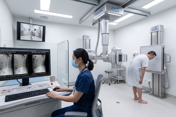
Operating dynamic dr safely requires that every examination begins with clinical necessity. Equipment should never be activated for exploratory screenings without documented diagnostic questions.
Clinical teams must exercise extra vigilance with radiosensitive populations, including pediatric patients and individuals who may be pregnant. In these cases, clinicians evaluate whether non-ionizing modalities can answer the diagnostic question before proceeding with dynamic digital radiography. When an examination is medically justified, certified radiologic technologists must oversee the process, ensuring beam filtration is active and patient position remains stable throughout the movement sequence. Adhering to institutional safety guidelines and local radiation safety legislation protects patients from avoidable diagnostic exposure.
Safety Features Buyers Should Prioritize in Dynamic Digital Radiography
Procurement specialists and clinical directors evaluating dynamic dr systems should focus on hardware and software controls that streamline dose governance.
A reliable system must feature responsive pulsed exposure controls that let operators fine-tune frame counts and pulse widths with precision. Built-in dose tracking displays, automated dose area product calculation, and structured DICOM dose reporting tools are equally indispensable for hospital audits. Beyond the hardware console, facilities should assess the vendor’s operator training resources, routine calibration protocols, and ongoing quality assurance support. A well-engineered unit operated by thoroughly trained staff remains the surest path to safe diagnostic imaging.
Conclusion
Dynamic digital radiography delivers safe clinical performance when teams combine appropriate medical indications with optimized exposure parameters and disciplined operational workflows. The technology should never be mischaracterized as entirely free from radiation risk, as it relies directly on diagnostic X-rays. Maintaining high safety standards requires healthcare facilities to pair modern pulsed hardware with conscientious technique, thoughtful protocol customization, and comprehensive technologist training across daily clinical routines.
Contact our technical imaging team today to explore radiation safety features and review dynamic digital radiography systems engineered for your clinical practice.