
What Are the Disadvantages of Digital Mammography?
Understanding the Limitations of Digital Mammography
Digital Mammography has changed the way healthcare providers perform breast cancer screening. Unlike traditional film mammography, it uses digital detectors to capture breast images and allows doctors to adjust, store, and review images more efficiently.
The technology offers many advantages. It provides faster image access, better image management, and improved workflow in hospitals and screening centers. However, Digital Mammography also has some limitations that patients and healthcare providers should understand.
A digital mammogram is not a perfect diagnostic method. It can detect many breast abnormalities, but some cancers or suspicious areas may still be difficult to identify. Factors such as breast density, image quality, and interpretation experience can affect the final results.
Understanding the disadvantages of digital mammography does not mean questioning its value. Instead, it helps patients understand what the technology can provide and when additional examinations may be needed.
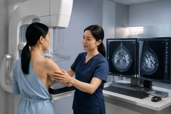
Possible Limitations in Detecting Breast Abnormalities
One of the main limitations of digital mammography is that it may not detect every abnormal change in breast tissue.
Small tumors, early-stage cancers, or abnormal areas with similar characteristics to normal breast tissue can sometimes be difficult to identify. Even though digital technology improves image processing and allows radiologists to adjust contrast and brightness, it cannot completely remove all detection challenges.
The accuracy of a digital mammogram depends on several factors. Breast structure, image quality, equipment performance, and the experience of the radiologist all influence the interpretation process.
Compared with traditional film mammography, Digital Mammography provides more flexibility in image review. Radiologists can enlarge images and adjust specific areas for closer evaluation. However, better image processing does not guarantee that every abnormality will be visible.
For this reason, doctors may combine mammography results with physical examinations, medical history, or additional breast imaging when necessary.
Dense Breasts Can Affect Mammogram Accuracy
Breast density is one of the most important factors affecting digital mammogram accuracy.
Dense breast tissue contains more glandular and fibrous tissue and less fatty tissue. On a mammogram, dense tissue appears white. The challenge is that many breast cancers also appear as white areas. This similarity can create a masking effect, making some abnormalities harder to distinguish.
Women with dense breasts may have a higher chance of receiving additional imaging recommendations after a mammogram. This does not always mean that cancer is present. It means that the doctor may need more information to evaluate the breast tissue clearly.
Additional examinations, such as ultrasound or MRI, may sometimes be used together with Digital Mammography. These imaging methods provide different information and can help doctors evaluate areas that are difficult to assess on a mammogram alone.
Because breast density varies among individuals, healthcare providers consider it an important factor when planning breast cancer screening strategies.
Radiation Exposure and Safety Considerations
Since Digital Mammography uses X-rays to create breast images, patients are exposed to a small amount of radiation during the examination.
Radiation exposure is a common concern among patients. However, modern Digital Mammography systems are designed to use controlled radiation doses while maintaining image quality. For most women, the benefits of early breast cancer detection are considered greater than the potential risks from this low-level exposure.
Repeated screening examinations over many years do result in cumulative radiation exposure. Therefore, healthcare providers usually recommend screening schedules based on age, risk factors, and clinical guidelines.
The goal of mammography is not to eliminate all risks but to provide an effective balance between early detection and patient safety.
Breast Compression May Cause Temporary Discomfort
During a Digital Mammography examination, breast compression is required to obtain clear images.
The breast is positioned between two plates and compressed for a short period while images are captured. Some patients may experience pressure, discomfort, or temporary pain during this process.
Although compression can be uncomfortable, it is an important part of breast imaging. It helps spread breast tissue evenly, reduces tissue overlap, minimizes motion blur, and allows the system to produce clearer images with a lower radiation dose.
The level of discomfort differs between individuals. Some patients may experience only mild pressure, while others may find compression more uncomfortable. Communicating with the imaging technician can help improve the examination experience.
Higher Equipment Cost and Maintenance Requirements
For hospitals and medical facilities, another disadvantage of Digital Mammography is the cost of equipment investment and maintenance.
Digital Mammography systems require advanced detectors, image processing software, and regular quality control procedures. Compared with traditional film-based systems, the initial purchase cost can be higher.
Healthcare facilities also need trained technicians and technical support to maintain system performance. Regular calibration, software updates, and equipment inspections are necessary to ensure consistent image quality.
For medical organizations planning to upgrade their breast imaging services, equipment cost is an important consideration. The decision usually involves balancing investment requirements with long-term benefits, including improved workflow and digital image management.
Professional Interpretation Remains Essential
Although Digital Mammography improves image quality, the final interpretation still depends on trained healthcare professionals.
Radiologists need experience to identify subtle changes and determine whether further evaluation is required. Different interpretations may occur depending on image appearance and clinical information.
Digital Mammography can sometimes lead to false-positive results, where a suspicious finding turns out to be harmless after additional testing. This may result in extra imaging examinations or biopsies.
False-negative results can also occur when an abnormality is present but not visible on the mammogram. These possibilities show why professional interpretation and appropriate follow-up are important parts of breast cancer screening.
Conclusion: Understanding the Role of Digital Mammography
Digital Mammography remains one of the most important tools for breast cancer screening. It provides significant improvements compared with traditional film mammography and supports more efficient breast imaging workflows.
However, understanding the disadvantages of digital mammography is also important. Limitations related to dense breasts, radiation exposure, breast compression, equipment costs, and image interpretation should be considered when evaluating this technology.
Digital Mammography is not designed to replace every other breast imaging method. Instead, it serves as a reliable screening tool that works together with clinical evaluation and additional imaging when needed. By understanding both its benefits and limitations, patients and healthcare providers can make better decisions about breast health management.

