
What’s the Difference Between a Regular Mammogram and a Digital Mammogram?
Mammography has been widely used for breast cancer screening and diagnosis for decades. However, the technology behind mammography has changed significantly. Traditional film-based systems have gradually been replaced by digital solutions that improve how breast images are captured, processed, and managed.The main difference between a regular mammogram and a Digital Mammogram is not the X-ray principle itself. Both methods use low-dose X-rays to create images of breast tissue. The difference lies in how the X-ray information is recorded after it passes through the body.
A regular mammogram uses film to capture and store the image, while a Digital Mammogram uses electronic detectors to convert X-ray signals into digital data. This change has transformed breast imaging workflows and provided radiologists with more flexibility during image review.
Traditional Mammogram: Film-Based Breast Imaging
A regular mammogram typically refers to conventional film-screen mammography. In this method, X-rays pass through compressed breast tissue and expose a film-based image receptor.
The film records differences in X-ray absorption between different areas of the breast. After exposure, the film must undergo chemical processing before the image can be viewed.
Film mammography was an important development in breast cancer screening because it allowed clinicians to detect abnormalities that could not be identified through physical examination alone. However, this technology also had limitations.
One major limitation was that the image appearance was fixed after exposure. If exposure conditions were not ideal, important details could become difficult to visualize. Adjustments to brightness or contrast were not possible after the film was processed.
Film storage was another challenge. Healthcare facilities needed physical space to store patient images, and sharing previous examinations between different locations could be inconvenient.
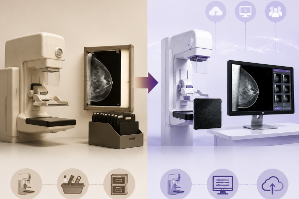
Digital Mammogram: A Digital Imaging Workflow
A Digital Mammogram replaces film with an electronic detector system. Instead of creating a physical image, the system captures X-ray signals digitally and processes them through specialized software.
This approach is a specialized application of Digital Radiography. The detector receives X-ray information after it passes through breast tissue and converts it into electronic signals.
The original image data can then be processed, displayed, and stored electronically. Radiologists can adjust image presentation during review, allowing them to examine different areas of breast tissue more effectively.
Modern Mammography Digital Radiography systems are designed specifically for breast imaging, where small differences in tissue structure may have clinical importance.
Image Capture Technology: Film Compared with Digital Detector
The most fundamental difference between a regular mammogram and a Digital Mammogram is the image receptor and the way image data is collected.
Traditional mammography uses film as the recording medium. The film directly captures the X-ray exposure pattern, but the available image information is limited by the characteristics of the film.
A Digital Mammogram uses advanced detectors, including Flat Panel Detector technology in many modern systems. These detectors convert X-ray photons into electrical signals and preserve the information as digital data.
This digital acquisition method provides greater flexibility because the captured information can be processed after exposure. It also allows manufacturers to optimize detector sensitivity, resolution, and noise performance for breast imaging applications.
Because breast abnormalities can be very subtle, detector performance plays an important role in maintaining image detail and contrast.
Image Processing and Display Differences
Another important difference between a regular mammogram and a Digital Mammogram is how images are processed after acquisition.
With film mammography, the final image appearance is determined during exposure and film development. If the image contrast or exposure is not suitable, repeating the examination may be necessary.
Digital Mammography separates image acquisition from image display. After the detector captures the X-ray data, image processing software analyzes and optimizes the information before the image is presented to the radiologist.
This process allows adjustments to contrast, brightness, and noise reduction. These capabilities are especially useful in breast imaging because radiologists often need to evaluate small structures, such as microcalcifications or subtle density changes.
However, image processing cannot create information that was not captured by the detector. The quality of the original digital data remains essential for accurate image interpretation.
Storage and Workflow in Modern Breast Imaging
The way images are stored and shared is another major difference between a regular mammogram and a Digital Mammogram.
Film-based mammograms require physical storage and manual management. Reviewing previous examinations often requires locating stored films and transporting them between departments or facilities.
A Digital Mammogram creates electronic image files that can be integrated into modern medical imaging networks. Images can be stored in systems such as PACS, accessed by authorized healthcare professionals, and compared with previous examinations more efficiently.
For hospitals and diagnostic centers, digital workflow improves accessibility and supports faster communication between radiologists and clinicians.
Compared with traditional film-based methods, Digital Mammography provides a more efficient approach for managing large numbers of examinations. This is especially important for modern breast cancer screening programs, where image accessibility and workflow efficiency directly affect clinical operations.
Diagnostic Flexibility of Digital Mammography
Both traditional and digital mammograms can be used for breast cancer screening and diagnostic evaluation. However, a Digital Mammogram provides greater flexibility in modern clinical environments.
For screening programs, digital systems support efficient image management when large numbers of examinations are performed. Images can be reviewed, stored, and compared more easily.
For diagnostic mammography, digital images allow radiologists to adjust image display and focus on specific areas that require further evaluation.
Digital technology also provides a foundation for advanced applications, including computer-assisted analysis and artificial intelligence-based image evaluation.
The improved flexibility of a Digital Mammogram does not come from changing the basic X-ray imaging principle. Instead, it comes from improvements in image acquisition, processing, storage, and clinical workflow.
The Role of Digital Mammography in Modern Breast Imaging
The transition from film-based mammography to digital imaging represents more than a change in image storage. It reflects a broader transformation toward connected, data-driven healthcare.
Modern Digital Mammography systems combine detector technology, image processing, and digital workflow management to support more efficient breast imaging.
The development of Flat Panel Detector technology has further improved image acquisition by providing better signal conversion and consistent performance across different examinations.
As healthcare facilities continue to upgrade their imaging infrastructure, digital mammography has become an important component of modern breast cancer screening and diagnostic services.
In comparison with a regular mammogram, a Digital Mammogram offers improved flexibility in handling image data and supports the requirements of modern medical imaging environments.
Digital Mammogram and Modern Imaging Technology
A Digital Mammogram is an important example of how digital technologies have changed traditional X-ray imaging. Similar to other Digital X-ray applications, it converts captured X-ray information into electronic data instead of relying on physical film.
The combination of detector technology, software processing, and digital storage allows healthcare providers to manage images more effectively.
As a specialized form of Digital Radiography, Mammography Digital Radiography focuses on the unique requirements of breast imaging. It requires high image quality, accurate contrast representation, and reliable system performance.
These improvements help radiologists review images more efficiently and support better clinical decision-making during screening and diagnosis.
Conclusion
The difference between a regular mammogram and a Digital Mammogram mainly comes from how image information is captured, processed, and managed.
Traditional mammography relies on film to record X-ray images, while a Digital Mammogram uses electronic detectors and digital image processing technologies to create, display, and store image data.
By combining Flat Panel Detector technology with advanced software, digital systems provide greater flexibility for image review, storage, and clinical workflow.
The development of Digital Mammography has improved the efficiency of modern breast imaging and has become an important part of breast cancer screening and diagnostic services.
At SEEHO Medical Equipment, we focus on medical imaging technologies that support modern healthcare environments. Our Mammography Digital Radiography systems are designed to provide reliable breast imaging performance for hospitals, diagnostic centers, and healthcare providers seeking advanced digital imaging solutions. If you are planning to purchase a new Digital X-ray Machine or upgrade an existing imaging device, welcome to contact us. Our professional team will provide suitable product solutions and technical support.

