
Digital Mammography vs. Breast MRI: When is Each Modality Recommended?
Digital mammography serves as the universal first-line tool for routine breast cancer screening, while breast MRI acts as a specialized, high-sensitivity supplemental modality for high-risk patients and complex diagnostic assessments. In modern clinical practice, these two imaging technologies do not compete directly; rather, they complement each other to provide comprehensive diagnostic coverage across diverse patient risk profiles.
Healthcare facilities, diagnostic imaging centers, and outpatient clinics must understand how to deploy each modality effectively. Evaluating clinical indications, acquisition physics, equipment footprint, and operational overhead helps administrators optimize patient care and capital allocation.
Key Differences in Imaging Technology and Purpose: Digital Mammography vs. Breast MRI
Understanding the physical principles behind each system clarifies their distinct roles in clinical diagnostic workflows.
How Digital Mammography Works as Primary Screening
Full-field digital mammography uses low-dose ionizing radiation with specialized digital flat-panel detectors (FPD). The system compresses breast tissue briefly to acquire high-resolution, two-dimensional projections.
This mechanism provides exceptional spatial resolution, making it the global standard for detecting subtle microcalcifications and early structural tissue distortions. Standard examinations require only a few minutes per patient, making digital mammography the primary foundation for high-throughput screening programs.
How Breast MRI Works for High-Risk Assessment
Breast Magnetic Resonance Imaging (MRI) relies on strong magnetic fields, radiofrequency pulses, and intravenous gadolinium-based contrast agents rather than ionizing radiation.
The contrast agent highlights areas of increased blood supply (neoangiogenesis), which frequently characterizes malignant tumors. This functional imaging technique produces multi-planar, cross-sectional slices that offer outstanding soft-tissue contrast, allowing clinicians to evaluate the exact vascular morphology and tumor extent in three dimensions.
Clinical Indications in Digital Mammography vs. Breast MRI
Patient risk factors, clinical history, and anatomical tissue density dictate when each examination method is appropriate.
When Digital Mammography is the First-Line Standard
Digital mammography remains the standard initial examination for the vast majority of women:
- Routine Population Screening: Recommended for asymptomatic women starting at age 40 to identify early subclinical abnormalities.
- Microcalcification Detection: Superior spatial clarity makes digital X-ray imaging the most reliable method to visualize clustered microcalcifications associated with ductal carcinoma in situ (DCIS).
- Initial Symptom Workup: Ideal first-line diagnostic evaluation for palpable lumps, localized pain, or nipple discharge before escalating to advanced cross-sectional modalities.
When Contrast-Enhanced Breast MRI is Required
Breast MRI is reserved for specific, highly targeted diagnostic circumstances:
- High Genetic or Familial Risk: Women with confirmed BRCA1 or BRCA2 gene mutations, or those with a lifetime risk exceeding 20%, benefit from annual supplemental MRI.
- Occult Primary Evaluation: Detecting hidden primary lesions when metastatic breast cancer is diagnosed in axillary lymph nodes without an obvious mammographic finding.
- Preoperative Staging: Mapping tumor boundaries, multifocal disease, and chest wall involvement prior to breast-conserving surgery.
- Dense Breast Assessment: Evaluating suspected lesions in women with extremely dense breast tissue when baseline radiographic and ultrasound findings remain inconclusive.
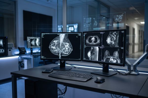
Facility Cost, Workflow, and Accessibility Comparison
Operating these imaging systems presents vastly different technical, physical, and financial demands for healthcare providers.
| Operational Factor | Digital Mammography System | Dedicated Breast MRI Setup |
| Initial Capital Expenditure | Accessible, budget-friendly for clinics | Extremely high capital investment |
| Room & Shielding Requirements | Standard lead shielding, compact space | RF-shielded room, heavy structural floor |
| Exam Duration | 3 to 5 minutes per patient | 30 to 45 minutes per patient |
| Daily Patient Throughput | High throughput (20–40+ exams/day) | Low throughput (6–10 exams/day) |
| Contrast Agent Dependency | None (non-invasive, low dose) | Requires IV contrast (gadolinium) |
| Operational Complexity | Standard radiographic technician | Specialized MRI technologist & nurse |
Equipment Footprint and Shielding Requirements
A digital mammography unit features a compact, floor-mounted design that integrates smoothly into standard diagnostic examination rooms. Structural requirements involve conventional lead lining in walls and doors to manage low-energy scatter radiation.
In contrast, an MRI installation demands extensive space planning, dedicated equipment rooms, cooling systems (chillers and cryogen management), and specialized copper radiofrequency (RF) shielding to prevent external signal interference.
Scan Duration and Patient Throughput
Workflow speed directly impacts operational sustainability and patient scheduling. A routine bilateral digital mammography examination takes less than 5 minutes from positioning to exposure completion.
A contrast-enhanced breast MRI procedure generally spans 30 to 45 minutes. The patient must remain prone and motionless inside the scanner bore, requiring IV line placement, contrast injection protocols, and extended acquisition sequences.
Building an Efficient Breast Diagnostic Strategy
A balanced imaging center organizes its clinical capabilities to match local healthcare demand and budget constraints.
Primary Screening vs. Supplemental Diagnostic Modalities
For private clinics, community healthcare facilities, and regional diagnostic centers, establishing a high-performance digital mammography unit is the primary operational priority. It handles the volume of routine preventive health checks and provides rapid, actionable findings.
Facilities can then build referral networks with specialized tertiary hospitals for supplemental breast MRI services when complex, high-risk cases arise. This tiered approach optimizes facility resources without compromising diagnostic pathways.
Practical Takeaways for Imaging Center Planning
Digital mammography provides the frontline speed, spatial resolution, and affordability needed for widespread preventive screening. Breast MRI delivers deep soft-tissue sensitivity for high-risk surveillance and detailed surgical staging at a much higher operational cost. Aligning your equipment selection with patient demographics and workflow volume ensures clinical precision and steady financial performance.
If you are evaluating new digital mammography systems or planning to upgrade your outpatient radiology suite, contact our technical team for detailed product specifications and facility layout solutions.

