
2D vs 3D Digital Mammography: Which System Fits Your Facility’s Budget?
For small to mid-sized clinics and outpatient centers, standard 2D digital mammography offers the best balance of clinical reliability, lower upfront cost, and rapid financial return. While 3D digital mammography (tomosynthesis) provides multi-slice imaging for dense breast tissue, it requires significantly higher initial capital, larger PACS storage, and ongoing software maintenance.
With the rising global demand for early breast cancer screening, healthcare providers must evaluate their clinical goals against real equipment budgets. Understanding the operational differences between these two modalities ensures your facility makes a sound financial decision.
Understanding the Core Technology Difference
Evaluating digital breast imaging requires a clear look at how each modality captures diagnostic data.
Standard 2D Digital Mammography (FFDM)
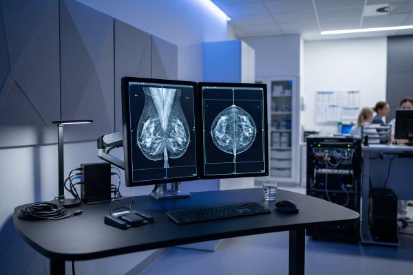
Full-Field Digital Mammography (FFDM) captures two-dimensional X-ray images of the compressed breast from two standard angles: cranio-caudal (CC) and mediolateral oblique (MLO). The X-ray beam passes through the tissue directly onto a high-efficiency flat-panel detector.
This configuration produces clear, high-contrast digital images ideal for identifying microcalcifications and distinct structural masses. Because the imaging process involves static exposures, exposure times remain brief, and data processing occurs almost instantaneously.
3D Breast Tomosynthesis (DBT)
Digital Breast Tomosynthesis (DBT) moves the X-ray tube in an arc across the breast, capturing multiple low-dose projection images at various angles. Specialized reconstruction algorithms compile these projections into a series of thin, high-resolution 1-millimeter cross-sectional slices.
This slicing technique minimizes tissue overlap, making it easier for radiologists to inspect dense fibroglandular tissue. However, this advanced acquisition method requires complex gantry engineering, higher processing power, and specialized viewing software.
Cost and Financial Breakdown in 2D vs 3D Digital Mammography
The financial commitment for breast imaging extends well beyond the initial purchase order.
Upfront Equipment and Installation Expenses
Initial capital expenditure represents the largest immediate cost difference. A reliable 2D digital mammography system generally requires a much lower upfront investment. This makes it an accessible upgrade for private practices and community clinics.
In contrast, a 3D DBT unit involves precision motorized motion components, advanced reconstruction hardware, and specialized review workstations. These elements can increase baseline acquisition costs significantly. Furthermore, DBT systems often demand enhanced structural site planning and dedicated high-performance workstations for reading physicians.
PACS Storage and Network Bandwidth Demands
A standard 2D digital mammography examination produces a lightweight dataset of four standard DICOM images. These files transfer quickly across existing local networks and require minimal storage on Picture Archiving and Communication Systems (PACS).
A single 3D tomosynthesis exam can generate dozens or hundreds of image slices, resulting in substantial file sizes. Facilities investing in 3D systems must frequently upgrade network bandwidth, purchase expanded server storage, and budget for ongoing cloud archival costs.
Clinical Throughput and Return on Investment (ROI)
Clinical daily workflow directly governs how quickly an imaging system pays for itself.
Exam Speed and Daily Patient Volume
Patient throughput is a critical factor for facilities operating in high-demand or resource-conscious settings. Standard 2D digital systems complete examinations within a few minutes per patient. Fast exposure times and rapid image rendering allow radiographers to maintain a smooth, uninterrupted schedule.
While 3D units provide detailed image sets, the scanning pass and data reconstruction take slightly longer per exposure. Technicians must also manage patient positioning carefully during the extended arc movement.
Maintenance and Long-Term Operating Costs
Both imaging modalities require routine quality assurance and preventive care. However, the mechanical simplicity of a 2D floor-mounted system reduces wear on moving parts. Replacement X-ray tubes, detector maintenance, and annual software licensing for 2D units remain budget-friendly.
With 3D systems, ongoing service contracts and proprietary software licensing tend to be higher, requiring consistent daily patient volume to offset overhead.
How to Choose the Right Option for Your Facility
Selecting an imaging system depends on your target patient demographic, diagnostic scope, and capital structure.
When 2D Digital Mammography is the Ideal Choice
A 2D digital mammography system represents the most cost-effective option for:
- Private outpatient clinics and community health centers establishing a new breast screening service.
- Healthcare institutions transitioning from traditional analog film or CR equipment to fully digital workflows.
- Facilities prioritizing rapid patient throughput, straightforward equipment maintenance, and a fast breakeven timeline.
When Investing in 3D Makes Clinical and Business Sense
A 3D tomosynthesis system is better suited for:
- Specialized comprehensive oncology centers and tertiary hospitals handling high-risk referrals.
- High-volume diagnostic facilities where reduced patient recall rates for dense breast tissue justify greater capital expenditure.
- Institutions equipped with robust IT infrastructure and dedicated radiology IT support.
Practical Takeaways for Equipment Procurement
Both 2D full-field systems and 3D tomosynthesis units serve vital roles in modern preventive healthcare. Standard 2D digital mammography continues to deliver dependable diagnostic accuracy, high throughput, and manageable maintenance overhead for mid-volume facilities. Advanced 3D systems offer multi-slice visualization for complex clinical cases at a higher total cost of ownership. Aligning equipment capabilities with your daily exam numbers ensures sustainable clinical performance.
If you are planning to upgrade your diagnostic imaging department or add new breast screening capabilities, feel free to contact our team for tailored equipment configurations and technical specifications.

