
Which Is Better, a Digital or 3D Mammogram?
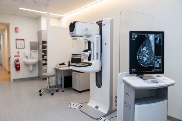
For the vast majority of outpatient clinics, diagnostic centers, and community screening programs, standard digital mammography remains the undisputed gold standard. While 3D breast tomosynthesis has emerged as a specialized multi-slice technology, standard 2D digital mammography offers superior spatial resolution for early calcification detection, lightning-fast patient throughput, and unmatched cost-efficiency.
Understanding why standard digital imaging continues to anchor modern breast screening helps healthcare providers choose equipment that balances high diagnostic reliability with rapid financial return on investment.
Detection Accuracy: Why Standard Digital Mammography Leads Routine Screening
Diagnostic success relies on spatial clarity and the rapid identification of subclinical lesions before they progress.
Unmatched Microcalcification Detection with 2D Systems
Standard 2D digital mammography utilizes high-resolution digital flat-panel detectors (FPD) that capture static, high-contrast X-ray projections. Because the detector does not move during exposure, it achieves exceptional pixel sharpness.
This makes 2D digital systems the superior modality for identifying clustered microcalcifications—often the very first sign of ductal carcinoma in situ (DCIS). For the majority of patients without extremely dense tissue, standard digital imaging provides clear, conclusive, and legally recognized diagnostic proof in seconds.
The Realistic Role and Limitations of 3D Tomosynthesis
3D tomosynthesis moves the X-ray tube across an arc to reconstruct multi-slice images. While this helps separate overlapping tissue in women with severely dense breast tissue, it introduces mechanical complexity and image reconstruction artifacts.
For general preventive screening, the incremental diagnostic gain of 3D rarely justifies the substantial increase in equipment acquisition, software licensing, and operational overhead.
Workflow, Patient Experience, and Radiation Safety
Operational efficiency and patient comfort are critical metrics for any busy radiology department.
| Clinical & Operational Factor | 2D Digital Mammography System | 3D Breast Tomosynthesis (DBT) |
| Microcalcification Sharpness | Superior spatial resolution (stationary exposure) | Can blur tiny calcifications during arc sweep |
| Compression & Scan Time | Fast (2–4 seconds per projection) | Extended (10–25 seconds continuous hold) |
| Patient Radiation Dose | Minimal baseline dose ($1\text{–}2\text{ mGy}$) | Higher total radiation exposure |
| Patient Daily Throughput | High capacity (30–50+ exams/day) | Slower turnover (10–15 exams/day) |
| PACS Bandwidth & Storage | Lightweight DICOM files (~50 MB/study) | Massive datasets (1–3 GB/study) |
| Equipment & Maintenance Cost | Cost-effective, accessible, fast ROI | Prohibitive purchase & service contract costs |
Minimal Radiation Exposure and Superior Patient Comfort
Standard 2D examinations deliver an exceptionally low radiation dose, fully compliant with international safety benchmarks. Furthermore, compression is applied only for a split-second exposure, releasing the breast immediately.
In contrast, 3D systems require patients to endure prolonged motorized compression while the tube completes its mechanical trajectory, increasing discomfort and patient movement artifacts.
Diagnostic Reliability and Universal Protocol Compliance
Decades of clinical trials validate standard 2D digital mammography as the global benchmark for mortality reduction.
Standard 2D images integrate effortlessly into established diagnostic workflows and universal BI-RADS reporting standards. They allow radiologists to make rapid, definitive comparisons against prior annual screening archives without navigating hundreds of reconstructed slice layers.
Economic Value: Maximizing Clinic ROI with Digital Mammography
From a financial and operational standpoint, standard digital units offer overwhelming practical advantages for private providers.
Lower Total Cost of Ownership and Infrastructure Demands
Purchasing and maintaining a 3D unit requires substantial capital, expensive proprietary tube replacements, and massive PACS server expansions to store heavy data stacks.
A high-performance 2D digital mammography unit integrates into standard X-ray suites with minimal lead shielding adjustments. It generates lightweight, standard DICOM files that transmit easily across local networks and tele-radiology clouds without costly bandwidth upgrades.
High Patient Throughput for Community Programs
Private practices and screening outreach programs thrive on smooth patient flow. With rapid positioning cycles and near-instant image preview, a 2D digital mammography system allows a single technician to screen 30 to 50 patients per day, ensuring rapid equipment payback and steady revenue generation.
Practical Takeaways for Imaging Center Planning
When asking which technology is better, the practical answer is clear: standard digital mammography provides the optimal combination of clinical precision, low patient dose, rapid workflow, and economic viability. While 3D systems remain a specialized niche for high-risk diagnostic workups, 2D FFDM is the reliable backbone of modern preventive healthcare.
If your facility is upgrading its radiology suite or seeking a dependable, high-yield digital mammography system, contact our technical specialists today for customized equipment configurations and ROI consultations.

