
Is a Digital Mammogram Less Painful?
Fear of physical discomfort remains one of the primary reasons women delay routine breast cancer screening. Patients frequently ask: is a digital mammogram less painful than traditional screening methods? The straightforward answer is yes. Advances in modern digital mammogram engineering have significantly transformed the patient experience through intelligent motorized pressure regulation, ergonomic curved compression paddles, and ultra-fast exposure speeds.
Understanding how current digital imaging technology balances necessary anatomical flattening with physical comfort helps healthcare providers ease patient anxiety and improve screening compliance.
Why Compression is Necessary During a Digital Mammogram
Although breast compression is often associated with discomfort, it serves vital physical and clinical functions in radiological imaging.
Spreading Tissue for Diagnostic Clarity
Adequate compression gently flattens the overlapping layers of fibroglandular structures. By spreading the tissue evenly across the image receptor, the system prevents normal glandular shadows from masking small underlying lesions. This mechanical flattening optimizes image contrast, allowing radiologists to distinguish between benign glandular tissue and early suspicious masses with maximum confidence.
Reducing Radiation Dose and Motion Blur
Thinner anatomical tissue requires significantly less X-ray penetration. Proper compression lowers the overall radiation dose necessary for diagnostic image quality. Furthermore, firm stabilization immobilizes the breast, eliminating motion artifacts caused by patient breathing or muscle tension during exposure.
Technological Improvements in Modern Digital Mammogram Systems
Next-generation digital hardware has replaced rigid mechanical clamping with responsive, patient-friendly features.
| Comfort Feature | Traditional Screen-Film/CR | Modern Digital Mammography System |
| Compression Control | Manual mechanical clamping | Intelligent motorized pressure sensing |
| Paddle Geometry | Hard, flat, sharp-edged plates | Flexible, curved, ergonomic paddles |
| Exposure Duration | Slow ($3\text{–}6\text{ seconds}$ per view) | Instantaneous ($1\text{–}2\text{ seconds}$ per view) |
| Pressure Release | Manual lever release | Automatic, instant post-exposure release |
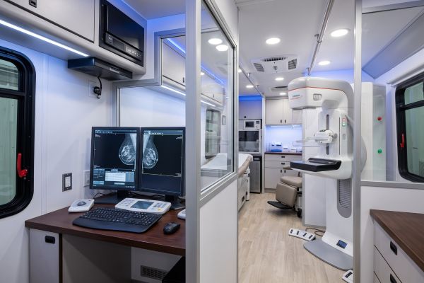
Smart Motorized Gentle Compression Controls
Contemporary digital units feature microprocessor-controlled compression systems. Instead of applying arbitrary force, pressure sensors dynamically gauge tissue resistance and stop advancing once optimal diagnostic thickness is reached. This automated control prevents unnecessary over-compression while ensuring compliant image quality.
Flexible and Ergonomic Paddle Designs
Unlike older flat, rigid plates that exerted uneven focal pressure on the chest wall, modern digital units utilize flexible, curved paddles. These paddles contour naturally to the breast’s anatomical shape, distributing force evenly across the surface and eliminating painful localized pinch points.
Faster Exposure Times with Digital Detectors
High-sensitivity amorphous silicon or selenium flat-panel detectors require only a fraction of a second to register ionizing photons. Because exposure is virtually instantaneous, the motorized paddle releases pressure immediately, reducing total compression time to just two to three seconds per projection.
Clinical Factors that Influence Patient Comfort
Individual anatomical differences and clinical technique also affect the examination sensation.
Breast Tissue Density and Sensitivity
Women with dense breast tissue or pre-existing fibrocystic conditions may experience greater tenderness during compression. Hormonal fluctuations during the menstrual cycle can also heighten breast sensitivity.
Proper Radiographer Positioning Techniques
An empathetic, skilled radiographer plays a crucial role in minimizing discomfort. Proper ergonomic positioning, smooth skin-smoothing motions, and gradual paddle descent keep the pectoral muscle relaxed, dramatically reducing procedural tension.
Practical Tips for Improving Patient Experience in Clinics
Healthcare facilities can implement practical operational workflows to ensure a calm, gentle examination:
- Schedule Around the Menstrual Cycle: Advise premenopausal patients to book appointments one to two weeks after their menstrual period ends, when breast tissue is least sensitive.
- Proactive Patient Communication: Instruct technologists to explain every step before applying compression, allowing patients to mentally prepare and relax upper body muscles.
- Warm and Welcoming Environment: Utilize ambient room warming and paddle-warming pads to prevent the shock of cold plastic on bare skin.
Combining Clinical Precision with Patient Comfort
Modern digital mammogram technology proves that diagnostic rigor does not have to come at the expense of patient comfort. With smart pressure-sensing controls, ergonomic contouring paddles, and split-second digital exposure times, today’s 2D digital systems offer a far gentler, faster, and more reassuring clinical experience than previous generations of equipment.
If you are planning to upgrade your radiology suite with an advanced, patient-centric digital mammography system, contact our technical team today for detailed equipment specifications and ergonomic design consultations.

