
How to Retrofit an Existing X-Ray Room for a New Digital Mammography System
Retrofitting a standard general radiography suite for a new digital mammography system is both structurally feasible and cost-effective because mammography operates at low tube voltages (20 to 40 kVp), requiring only 1.0 to 1.5 mm lead equivalence (Pb) for radiation safety. Unlike heavy CT or general X-ray rooms that demand 2.0 mm Pb or thicker barriers, the primary retrofit challenges center on providing adequate C-arm gantry clearance, installing dedicated electrical grounding, and maintaining strict HVAC climate control for delicate flat-panel detectors. Following a structured site planning process ensures clinical compliance while cutting renovation downtime.
Space Planning and Ergonomics for a Digital Mammography System
General radiography rooms usually offer ample square footage, but a dedicated mammography suite requires specialized floor layouts that balance mechanical clearance with patient privacy.
| Architectural Parameter | Standard General X-Ray Suite | Retrofitted Digital Mammography Suite |
| Minimum Floor Area | $16\text{ m}^2 – 20\text{ m}^2$ ($170 – 215\text{ sq ft}$) | $12\text{ m}^2 – 14\text{ m}^2$ ($130 – 150\text{ sq ft}$) |
| Clearance Radius | Linear ceiling-tube tracking paths | $360^{\circ}$ C-arm rotation plus wheelchair access |
| Shielding Requirement | $1.5\text{ mm} – 2.0\text{ mm Pb}$ (100–150 kVp) | $1.0\text{ mm} – 1.5\text{ mm Pb}$ (20–40 kVp) |
| Ambient Climate Spec | Standard hospital HVAC | $20^{\circ}\text{C} – 25^{\circ}\text{C}$ ($68^{\circ}\text{F} – 77^{\circ}\text{F}$), 30%–60% RH |
Minimum Room Dimensions and Gantry Clearance Requirements
A modern digital mammography system requires a minimum clear floor footprint of $3.5\text{ m} \times 3.5\text{ m}$ ($11.5\text{ ft} \times 11.5\text{ ft}$) with a ceiling clearance of at least $2.4\text{ m}$ ($8\text{ ft}$):
- Isocenter Rotation Arc: Allow a clear $1.5\text{ m}$ radius around the central column so the motorized C-arm can swing freely between CC (craniocaudal) and steep MLO (mediolateral oblique) angulations without colliding with side walls.
- Wheelchair Accessibility: Ensure wide entrance doors ($\ge 90\text{ cm}$) and unobstructed approach paths so technologists can lower the compression stand directly over seated patients.
- Service Clearances: Leave at least $60\text{ cm}$ behind the rear gantry housing to give field engineers unhindered access to high-voltage generators, circuit boards, and internal cooling pumps.
Private Changing Booth and Patient Flow Design
Patient anxiety directly influences muscle tension, which makes breast positioning harder and increases the risk of motion artifacts on a digital mammography system:
- Integrated Sub-Waiting and Changing Cabins: Build a direct-access dressing cubicle that opens straight into the scan room, eliminating exposure to public corridors.
- One-Way Traffic Separation: Where space permits, separate incoming patient lines from post-exam exit routes to speed up screening turnover.
- Acoustic and Visual Privacy: Add solid-core shielded doors and sound-dampening wall panels so patients feel comfortable discussing clinical history.
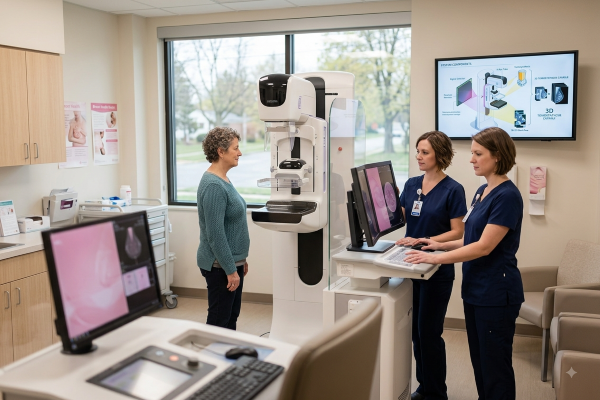
Structural and Radiation Shielding Requirements
Because mammography uses soft X-rays, existing general X-ray rooms almost always meet or exceed the primary structural barrier requirements.
Lead Equivalence Standards for Low-kVp Mammography (1.0–1.5 mm Pb)
General X-ray rooms are built with 1.5 mm to 2.0 mm Pb sheet lining, which easily blocks the low-energy scatter produced by a digital mammography system:
- Existing Wall Assessment: Have a qualified medical physicist conduct transmission surveys on existing drywall. In most cases, existing lead lining requires zero additional reinforcement.
- Doorway and Joint Flashing: Inspect wall seams, pass-through boxes, and service conduits. Any new electrical cutouts through existing lead walls must be backed with overlapping 1.5 mm Pb sheet patches.
- Floor and Ceiling Attenuation: Standard concrete slab floors ($\ge 10\text{ cm}$ thickness) offer sufficient intrinsic shielding for rooms located above occupied clinic spaces.
Control Console Lead Glass and Technician Viewing Angle
Technologist barrier placement requires direct, uninterrupted line-of-sight to both the patient and the gantry controls during the entire exposure sequence:
- Protective Barrier Rating: Install a fixed control screen with a lead-acrylic or lead-glass window certified to at least 1.0 mm Pb equivalence at 50 kVp.
- Viewing Angle Ergonomics: Position the console at a $45^{\circ}$ orientation toward the patient positioning zone so the operator can spot patient movement before triggering exposures.
- Safety Dead-Man Switch: Mount the secondary exposure switch behind the shielded zone, preventing technologists from initiating beams while standing outside the protected perimeter.
Electrical Infrastructure and Environmental Controls
Electronic flat-panel detectors (FPDs) are sensitive to power grid noise and ambient room temperature swings.
Power Supply, Dedicated Grounding, and Circuit Load Capacity
A digital mammography system demands clean, uninterrupted three-phase or single-phase utility power depending on generator sizing:
- Dedicated Distribution Panel: Run a dedicated line from the main hospital distribution board to avoid harmonic interference from heavy loads like elevators or MRI chillers.
- Low-Resistance Earth Grounding: Establish an isolated medical grounding bus with independent resistance below $2\,\Omega$ to eliminate image noise and micro-voltage spikes across the detector readout.
- Online Double-Conversion UPS: Place an online UPS between the wall breaker and the digital mammography system to stabilize voltage sags and provide emergency shutdown power during blackouts.
Dedicated HVAC and Continuous Dehumidification for the Detector
Amorphous Selenium (a-Se) and Cesium Iodide (CsI) detectors degrade when exposed to heat spikes and moisture:
- 24/7 HVAC Operation: Program climate control to maintain $20^{\circ}\text{C} – 25^{\circ}\text{C}$ continuously. Turning off AC over weekends causes thermal shock and permanent detector crystallization.
- Active Humidity Control: Maintain relative room humidity between 30% and 60% non-condensing to prevent static discharge on readout electronics.
Streamlining Your Clinical Facility Conversion
Repurposing an underutilized general X-ray suite for mammography avoids expensive structural additions while speeding up clinical deployment. Ensuring your room matches gantry rotation clearances, power stabilization specs, and environmental baselines guarantees artifact-free operation from day one.
Reach out to our facility planning and imaging specialists today to request architectural DWG layout templates, electrical load calculation guides, and customized site-readiness assessments for your digital mammography system.

