
Integrating AI Computer-Aided Detection (CAD) with digital mammography relies on standardized DICOM 3.0 handshakes to feed raw projection data into deep convolutional neural networks, delivering instant concurrent-reader assistance that flags occult microcalcifications and architectural distortions. Rather than replacing clinical judgment, modern algorithms process full-field acquisition data within milliseconds, scoring lesion malignancy probabilities and highlighting subtle soft-tissue masses before the radiologist opens the study. Understanding the shift from early rule-based systems to deep learning, along with local server architecture, helps imaging clinics optimize reading speeds, lower diagnostic recalls, and elevate overall screening accuracy.
The Evolution of CAD in Digital Mammography
Early computer-aided systems generated high false-positive marks that slowed reading workflows, but deep learning models have fundamentally changed algorithmic precision.
| Technical Parameter | Legacy Rule-Based CAD | Deep Learning AI CAD |
| Underlying Architecture | Hand-crafted edge/contrast filters | Multi-layer Convolutional Neural Networks (CNNs) |
| Lesion Recognition | Pixel threshold density mapping | Contextual morphological pattern recognition |
| False-Positive Prompts | 2.0 to 4.0 marks per normal case | Less than 0.5 prompts per normal case |
| Dense Breast Performance | Degraded by parenchymal overlap | Maintains high sensitivity via depth-feature extraction |
From Rule-Based Algorithms to Deep Learning Neural Networks
First-generation software applied rigid pixel-intensity formulas to standard digital mammography data, frequently tagging benign vascular calcifications and overlapping glandular tissue as suspicious lesions. Modern AI platforms train on millions of biopsy-proven screening cases. These neural networks evaluate structural asymmetry, border spiculation, and subtle tissue density gradients, adapting their sensitivity dynamically without flooding the reading monitor with distracting false prompts.
Automated Microcalcification and Mass Identification
Deep learning engines run specialized parallel analytical tracks to detect distinct breast pathologies:
- Microcalcification Cluster Analysis: Identifies individual specks down to 100 microns, analyzing cluster distribution, polymorphism, and spatial grouping patterns.
- Soft-Tissue Mass Segmentation: Penetrates fibroglandular backgrounds to map non-calcified masses, focal asymmetries, and architectural distortions that often hide behind dense parenchyma.
Clinical Benefits for Busy Imaging Centers and Clinics
Integrating AI algorithms directly into high-volume digital mammography reading rooms directly addresses reader fatigue while safeguarding diagnostic consistency.
Reducing Diagnostic False Negatives in Dense Tissue
Extremely dense breasts (BI-RADS Category C and D) present significant masking effects during regular screening:
- Parenchymal Penetration: AI models evaluate subtle parenchymal disruption around lesion margins, catching small invasive ductal carcinomas that human eyes might overlook during high-volume shifts.
- Consistent Diagnostic Floor: The neural network provides unwavering analytical sensitivity across an entire shift, preventing missed diagnoses caused by eye strain near the end of long reading lists.
Triaging High-Risk Cases and Alleviating Radiologist Fatigue
High-throughput breast screening centers handle hundreds of studies daily on each digital mammography workstation:
- Prioritized Worklists: AI engines pre-screen incoming DICOM studies in the background, automatically sorting priority cases with high malignancy risk scores to the top of the radiologist’s queue.
- Streamlined Normal Case Sign-Off: Clean screening cases with zero suspicious features are flagged for rapid review, cutting average per-case interpretation times without sacrificing screening accuracy.
Integrating AI Software into Your Digital Mammography Workflow
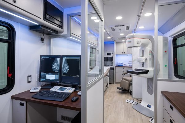
Successful software integration requires clean data pipelines between physical acquisition gantries, local PACS, and AI processing nodes.
Seamless DICOM 3.0 Handshake with Acquisition Workstations
An enterprise digital mammography suite connects AI engines via standard imaging protocols:
- Auto-Routing Configuration: The gantry console auto-forwards “For Processing” raw images straight to the AI server instantly after exposure.
- Secondary Capture & DICOM SR: The software generates standardized DICOM Structured Reports (SR) and transparent overlays that toggle on and off at the diagnostic workstation with a single keypress.
Cloud-Based vs. On-Premise AI Server Configurations
Imaging facilities choose deployment models based on internal IT bandwidth, patient data governance, and capital budgets:
- On-Premise GPU Appliances: Local AI servers sit behind the clinic firewall, processing studies in real time with zero external network reliance and zero recurring cloud bandwidth costs.
- Cloud-Native Processing: Suitable for distributed imaging chains, cloud architectures offload intensive GPU calculations to remote clusters, scaling compute power dynamically across multiple decentralized screening centers.
Future-Proofing Your Breast Screening Services
Pairing high-quantum-efficiency hardware with deep learning intelligence transforms standard diagnostic workflows into predictive, high-precision screening environments. Selecting hardware platforms that feature open integration architecture ensures your facility can easily adopt next-generation neural networks as diagnostic standards evolve.
Contact our imaging solutions engineers today to discuss open DICOM integration, evaluate high-resolution detector options, and explore an AI-ready digital mammography hardware setup for your clinical practice.

