
What Are the Key Features of a Modern Digital Mammography System?
Introduction
Breast imaging technology has changed significantly in recent years. Healthcare providers now expect imaging equipment to provide high-quality images, efficient workflows, and better patient experiences. A modern Digital Mammography System combines advanced hardware and software technologies to support breast cancer screening and diagnostic examinations.
Unlike traditional film-based mammography, digital systems capture images electronically and allow faster processing, storage, and review. These improvements help radiologists analyze breast tissue more effectively while allowing hospitals to manage imaging procedures more efficiently.
The performance of a Digital Mammography System depends on several key components, including detector technology, image processing, exposure control, workflow design, and digital connectivity.
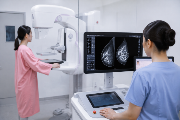
Advanced Imaging Technologies in Digital Mammography Systems
High-Resolution Detector
The detector is one of the most important components that determines image quality. During an examination, it captures X-ray signals and converts them into digital images for clinical evaluation.
A high-resolution detector allows a Digital Mammography System to display more detailed information from breast tissue. This capability is important when radiologists need to identify small calcifications, subtle tissue changes, or other potential abnormalities.
Detector performance also affects image consistency. In screening programs, healthcare facilities may perform many examinations every day. Stable image acquisition helps maintain reliable image quality across different patients and examination conditions.
Advanced Image Processing
After image acquisition, image processing software analyzes and optimizes the captured data. Modern processing technologies improve image contrast, reduce noise, and enhance important anatomical details.
For breast imaging, clear image presentation is essential because overlapping tissue structures can sometimes make interpretation more difficult. Advanced software helps radiologists review images with better visibility and supports more confident clinical decisions.
Image processing also helps balance image quality and radiation management. Instead of depending only on higher exposure levels, optimized software can improve image appearance through more efficient data processing.
Radiation Management and Patient Comfort Features
Automatic Exposure Control
Automatic Exposure Control (AEC) is a common feature in modern mammography equipment. It automatically adjusts exposure parameters based on breast thickness and tissue characteristics.
This function helps the Digital Mammography System maintain consistent image quality during different examinations. It reduces the need for manual adjustments and allows technicians to complete imaging procedures more efficiently.
Appropriate exposure control also supports patient safety by helping achieve diagnostic images while managing radiation dose.
Comfortable Compression and Positioning Design
Compression is an important step in mammography because it reduces tissue overlap and improves image accuracy. However, patient comfort has become an increasingly important consideration in modern system design.
Many advanced systems use improved compression mechanisms to provide more controlled pressure during examinations. Better compression design helps patients experience a more comfortable procedure while maintaining suitable imaging conditions.
Flexible positioning features also assist technicians during daily operation. Adjustable movements allow operators to position different patients more easily and improve examination efficiency.
Workflow Improvements in Digital Mammography Systems
Efficient Image Acquisition and Operation
A modern Digital Mammography System is designed to improve workflow efficiency from image acquisition to clinical review. Faster image capture allows technicians to complete examinations more smoothly, especially in busy screening centers.
User-friendly interfaces also simplify daily operation. Automated functions and clear controls help technicians manage patient positioning, image acquisition, and examination procedures with fewer complicated steps.
DICOM and PACS Integration
Digital connectivity is another important feature of current mammography solutions. DICOM compatibility allows imaging devices to exchange information with other medical systems using a standardized format.
PACS integration enables healthcare providers to store, access, and manage mammography images more efficiently. Radiologists can review previous examinations, compare image changes over time, and share information with authorized medical teams.
For hospitals building a digital imaging environment, connectivity capabilities are an essential part of a complete Digital Mammography System solution.
Conclusion
A modern Digital Mammography System combines imaging performance, workflow optimization, patient-focused design, and digital communication capabilities. Features such as high-resolution detectors, advanced image processing, automatic exposure control, and PACS connectivity help improve breast imaging procedures.
Understanding these key technologies allows healthcare providers to better evaluate different mammography solutions and select equipment that matches their clinical needs. Contact us to learn more about the advanced features of Digital Mammography Systems and find the right imaging solution for your facility.

