
Dynamic digital radiography: a novel quantitative modality to assess the pulmonary blood flow
Assessing pulmonary blood flow is important for understanding how the lungs function. Changes in blood circulation can affect oxygen exchange and may be associated with various respiratory and cardiovascular conditions. However, these changes are difficult to evaluate because conventional chest imaging mainly focuses on anatomical structures rather than physiological processes. Dynamic Digital Radiography (DDR) provides a new way to analyze these changes by capturing continuous X-ray images and evaluating variations related to pulmonary circulation.
Dynamic digital radiography (DDR) provides a new method for observing pulmonary circulation by analyzing continuous X-ray image sequences. Unlike traditional Digital Radiography, which captures a single image at a specific moment, DDR records time-dependent changes within the chest region. These changes can be further analyzed to obtain quantitative information related to pulmonary blood flow.
By combining dynamic image acquisition with quantitative analysis, Dynamic DR extends the role of X-ray imaging from structural observation toward functional assessment.
Understanding pulmonary blood flow changes through Dynamic digital radiography
Pulmonary blood flow changes continuously during the cardiac and respiratory cycles. The amount of blood reaching different regions of the lungs can influence X-ray attenuation, creating subtle variations between consecutive images.
Traditional chest radiographs can reveal conditions such as lung opacity changes or structural abnormalities, but they cannot directly show these dynamic circulation processes. More advanced imaging methods, including CT perfusion and nuclear medicine examinations, can provide functional information, but they may require more complex procedures.
Dynamic digital radiography captures multiple low-dose X-ray images in a short period. By analyzing the changes between these images, clinicians can observe variations related to pulmonary circulation and respiratory movement.
This time-based information provides a different perspective on lung assessment. Instead of evaluating only what the lungs look like, DDR helps investigate how lung regions change during physiological activity.
Quantitative analysis expands the clinical value of Dynamic digital radiography
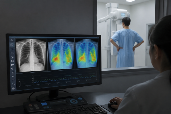
The key feature of Dynamic DR is not only continuous image capture but also the ability to extract measurable information from image sequences.
During pulmonary blood flow assessment, quantitative analysis methods can evaluate changes in pixel intensity over time. These variations may reflect differences in blood volume distribution within the lung fields. By processing these signals, researchers can create functional information that supports the evaluation of pulmonary circulation.
Compared with traditional radiographic interpretation, quantitative Dynamic DR analysis provides additional measurements rather than relying only on visual observation. This approach may help improve consistency when comparing different regions of the lungs or monitoring changes during follow-up examinations.
The combination of dynamic imaging and quantitative analysis makes DDR a potential tool for functional lung assessment.
Potential applications in pulmonary assessment
Dynamic digital radiography has been explored for evaluating pulmonary function and circulation-related changes. Its ability to capture real-time physiological processes makes it suitable for situations where anatomical images alone may not provide enough information.
For example, patients with pulmonary vascular abnormalities may show changes in blood distribution that are difficult to identify on conventional chest radiographs. DDR-based analysis may provide additional information about regional lung function and support clinical evaluation.
The technology may also be valuable in respiratory monitoring. By recording changes during breathing, DDR can provide information about both ventilation-related movement and circulation-related variations within the lungs.
Although Dynamic DR is still developing as a clinical tool, its combination of accessibility and functional analysis makes it an area of continued research in pulmonary imaging.
The future of functional lung imaging
Modern medical imaging is increasingly focused on combining anatomical information with functional assessment. Healthcare providers need imaging methods that can provide more clinical information while maintaining practical workflows.
Dynamic digital radiography offers a potential solution by adding quantitative analysis to a familiar X-ray imaging platform. Future improvements in image processing and artificial intelligence may further enhance the accuracy and efficiency of pulmonary blood flow assessment.
With continued research, Dynamic DR may become a complementary imaging method for evaluating lung function and supporting clinical decisions. It is not intended to replace CT, MRI, or nuclear medicine techniques, but to provide another option for functional assessment in selected clinical situations.
Conclusion
Pulmonary blood flow assessment requires information beyond traditional anatomical imaging. While conventional radiography remains an essential diagnostic method, it has limitations when evaluating dynamic physiological changes.
Dynamic digital radiography introduces a quantitative approach by analyzing continuous X-ray image sequences and extracting information related to pulmonary circulation. This combination of real-time imaging and data analysis provides a new perspective for functional lung evaluation.
For healthcare providers seeking advanced imaging solutions, SEEHO Medical Equipment offers reliable Digital Radiography technologies designed to support modern clinical workflows. With professional service and imaging solutions tailored to different healthcare needs, we help facilities improve diagnostic efficiency and patient care.

