
Dynamic Digital Radiography: Transforming Radiology Dynamics
Dynamic Digital Radiography (DDR) combines traditional X-ray imaging with real-time motion analysis, providing radiologists with additional information beyond static images. Unlike conventional Digital Radiography, which captures a single moment, this technology records a continuous sequence of images during movement and reveals how anatomical structures change over time.
X-ray imaging has always played an important role in medical diagnosis. For decades, radiologists have relied on Digital Radiography to evaluate anatomical structures such as bones, lungs, and soft tissues. However, some clinical conditions are related to functional changes rather than visible structural abnormalities.
Respiratory disorders may affect diaphragm movement, while musculoskeletal problems may influence joint motion during activity. In these situations, Dynamic Digital Radiography provides another way to evaluate patient conditions by showing both anatomical details and movement patterns.
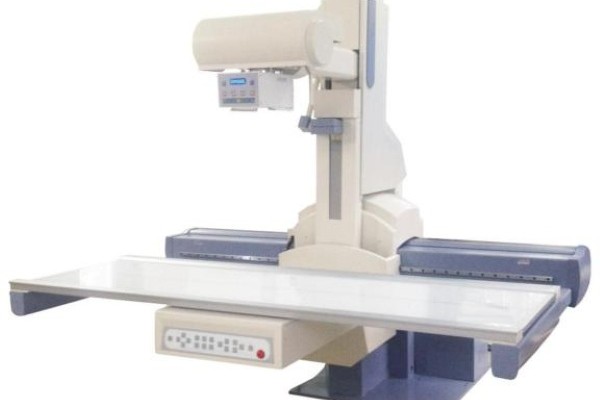
Moving Beyond Static X-ray Images with Dynamic Digital Radiography
Traditional Digital Radiography remains widely used because it is fast, accessible, and effective for many diagnostic tasks. However, static images have limitations when motion plays an important role in disease assessment.
A chest X-ray can show lung structure, but it cannot directly display how the diaphragm moves during breathing. A standard orthopedic radiograph can demonstrate bone position, but it may not show how a joint behaves during movement.
DDR helps address these limitations by capturing continuous image sequences. During the examination, patients can perform natural movements such as breathing or joint motion while the system records dynamic changes.
This additional information does not replace conventional X-ray imaging. Instead, it helps clinicians understand both the structure and function of the examined area.
Dynamic Digital Radiography in Clinical Evaluation
Dynamic Digital Radiography is gaining attention because many medical conditions involve changes that occur during movement.
In respiratory imaging, DDR can show diaphragm excursion and changes in lung expansion throughout the breathing cycle. These observations may provide additional information for patients with respiratory conditions where functional impairment is difficult to assess using static images alone.
The technology is also being studied in orthopedic applications. Joint instability and abnormal movement patterns may not always appear on a single radiograph. Dynamic sequences allow clinicians to observe motion patterns and compare functional changes during specific activities.
For radiologists, DDR provides another source of information when conventional imaging does not fully explain clinical symptoms. By combining structural images with motion analysis, clinicians can gain a more complete understanding of certain conditions.
Improving Radiology Workflow with Dynamic Digital Radiography
Radiology departments are under increasing pressure to provide accurate information while maintaining efficient workflows. Additional imaging examinations can increase patient waiting time and create extra workload for healthcare teams.
DDR provides dynamic information through a workflow similar to conventional Digital Radiography. Healthcare providers can collect motion-related data without introducing a completely different imaging process.
For example, a clinician evaluating respiratory function may review both anatomical images and diaphragm movement from the same examination. In orthopedic assessment, dynamic sequences can help evaluate joint mechanics alongside traditional structural findings.
By providing more information during one examination, DDR may help physicians make better-informed clinical decisions.
The Future of Dynamic Imaging in Radiology
The development of image analysis technology is expanding the possibilities of Dynamic DR. Reviewing a large number of sequential images can require additional time, especially when evaluating subtle movement changes.
Researchers are exploring automated analysis methods that can measure motion patterns, including diaphragm movement and joint displacement. These tools may help radiologists identify functional changes more efficiently.
Artificial intelligence may also become an important part of future DDR workflows. Instead of replacing radiologists, AI-based tools can assist with quantitative measurements and support image interpretation.
As these technologies continue to develop, dynamic imaging may become a more common part of radiology practice.
Conclusion
As radiology continues to develop, imaging technologies are expected to provide more detailed information while maintaining practical clinical workflows. DDR offers a way to extend the value of traditional X-ray examinations by adding motion-based evaluation to existing imaging procedures.
Although clinical applications are still expanding, current research shows that dynamic imaging can support a wider range of diagnostic needs. From respiratory evaluation to musculoskeletal assessment, DDR provides clinicians with additional information for understanding patient conditions.
SEEHO Medical Equipment offers advanced Digital Radiography solutions, including Dynamic DR systems designed to support modern diagnostic workflows. With reliable performance and professional service, we help healthcare providers select suitable imaging solutions for their clinical needs. Contact us for more information.

