
What Is Dynamic Range in Digital Radiography?
In Digital Radiography, image quality depends on more than spatial resolution and radiation dose. The ability of an imaging system to capture different levels of X-ray exposure is also an important factor. This capability is known as dynamic range.
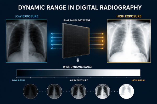
Dynamic range describes the range of X-ray signals that a detector can accurately record, from areas receiving very little radiation to regions exposed to higher levels of radiation. A wider dynamic range allows a Digital Radiography system to preserve more anatomical information within a single image.
Compared with traditional film-based radiography, modern DR Systems provide greater exposure flexibility. This reduces the influence of minor exposure variations and helps maintain consistent image quality across different examinations.
For hospitals evaluating a Digital X-ray Machine, dynamic range is an important technical factor because it influences image consistency, contrast representation, and diagnostic performance.
Understanding Dynamic Range in Digital Radiography
Dynamic range refers to the difference between the lowest and highest X-ray exposure levels that an imaging detector can capture while maintaining useful image information.
During an X-ray examination, different tissues absorb different amounts of radiation. Bones, soft tissues, and air-filled structures create various signal levels at the detector. The imaging system must accurately record these differences to produce a clear diagnostic image.
A detector with a limited dynamic range may lose information in extremely bright or dark areas. For example, highly exposed regions may become saturated, while low-exposure areas may show increased noise or reduced visibility.
Modern Digital Radiography systems overcome many of these limitations through advanced detector technology and image processing. These technologies allow the system to capture a wider range of exposure information before generating the final clinical image.
Dynamic Range Performance in Digital Radiography Systems
The performance of dynamic range is closely related to the design of the Digital Radiography system, especially the detector component.
The Flat Panel Detector (FPD) is responsible for receiving X-ray signals after they pass through the patient and converting them into digital data. The detector’s sensitivity, conversion efficiency, pixel structure, and electronic design all influence its ability to capture different exposure levels.
A high-performance Flat Panel Detector can preserve subtle differences between tissues while maintaining details in areas with stronger X-ray absorption.
For example, chest radiography contains both low-density lung regions and high-density bone structures. A detector with a suitable dynamic range can capture information from both areas within the same exposure, improving overall image quality.
Dynamic Range and Exposure Latitude
Dynamic range is often discussed together with exposure latitude, but these two concepts describe different aspects of imaging performance.
Dynamic range refers to the detector’s ability to record a wide range of X-ray signals. Exposure latitude describes how much variation in exposure conditions an imaging system can tolerate while still producing an acceptable image.
Traditional film-based radiography had a relatively narrow exposure latitude. Small exposure errors could significantly affect image brightness and contrast.
In comparison, modern Digital X-ray systems provide much greater flexibility. Image processing software can adjust brightness and contrast after image acquisition, allowing clinicians to obtain consistent images under different exposure conditions.
However, wider exposure latitude does not eliminate the need for proper dose control. Excessive exposure can increase patient radiation dose, while insufficient exposure may introduce image noise and reduce diagnostic value.
The Impact of Dynamic Range on Digital Radiography Imaging
Dynamic range has a direct influence on the performance of Digital Radiography imaging in different clinical applications.
Chest Imaging
Chest radiography is one of the most challenging examinations for dynamic range performance. The image contains structures with large differences in X-ray absorption.
The lungs, ribs, heart region, and soft tissues require different levels of image information. A wider dynamic range helps maintain visibility across these anatomical structures.
This allows radiologists to evaluate both lung details and surrounding structures within the same image.
Orthopedic Imaging
Orthopedic examinations require clear visualization of bone structures and fine anatomical details.
A Digital Radiography system with good dynamic range can capture dense bone areas while preserving information from surrounding soft tissues. This supports applications such as fracture assessment, joint evaluation, and postoperative monitoring.
Emergency Radiography
Emergency imaging often involves challenging conditions, including patient movement, positioning limitations, and different body sizes.
The wider exposure flexibility of modern DR Systems helps maintain stable image quality in urgent clinical situations where ideal imaging conditions may not always be possible.
Dynamic Range and Image Processing Technology
In Digital Radiography, the detector does not directly create the final displayed image. The captured signals are processed by software to optimize image appearance.
Image processing algorithms adjust parameters such as brightness, contrast, and noise reduction based on the acquired data. These technologies help convert a wide range of detector signals into clinically useful images.
However, image processing depends on the quality of the original data collected by the Flat Panel Detector. If important exposure information is not captured during acquisition, software cannot fully recover missing details.
Therefore, detector dynamic range and image processing technology work together to determine the final image quality of a DR System.
Dynamic Range in Modern DR System Selection
When selecting a Digital X-ray Machine, healthcare facilities usually consider multiple technical specifications, including image resolution, radiation efficiency, workflow, and detector performance.
Dynamic range is an important factor because it affects how consistently the system performs across different examinations and patient conditions.
A reliable DR System combines advanced Flat Panel Detector technology, optimized image processing, and effective exposure management. This combination helps healthcare providers achieve stable image quality while maintaining appropriate radiation control.
As Digital Radiography technology continues to develop, improvements in detector materials and artificial intelligence-based image processing may further enhance dynamic range performance.
Conclusion
Dynamic range is one of the key factors that influence the performance of a Digital Radiography system. A wider and more accurate exposure response helps maintain anatomical details across different examination types, from chest imaging to orthopedic assessment.
As DR technology continues to develop, detector performance, image processing capability, and system reliability will remain important considerations when selecting a Digital X-ray Machine.
At SEEHO Medical Equipment, we focus on providing reliable Digital Radiography systems and imaging solutions for healthcare facilities. If you are evaluating DR equipment for a new installation or system upgrade, our team can help you compare suitable configurations based on your imaging requirements and application scenarios.

