
Is Digital Mammography Suitable for Male Breast Cancer Diagnosis?
Many patients assume breast cancer develops exclusively in women. In reality, male breast cancer (MBC) accounts for roughly 1% of all breast cancer diagnoses worldwide.Public awareness regarding this disease remains remarkably low. Most healthcare systems do not provide routine screening programs for men. Consequently, male patients often ignore early clinical signs. They frequently overlook painless subareolar lumps and localized skin dimpling. These oversights cause men to receive clinical diagnoses at advanced stages. Delayed detection leads directly to poorer survival outcomes.Clinicians need dependable imaging tools to evaluate suspicious male chest masses promptly. In these clinical scenarios, digital mammography serves as the primary diagnostic foundation. This advanced modality provides the tissue contrast and spatial resolution required to evaluate male breast anatomy effectively.
Evaluating Gynecomastia and Diagnostic Accuracy With Mammographic Imaging
Clinicians must quickly distinguish benign tissue enlargement from invasive malignancies. Benign breast tissue proliferation is known as gynecomastia. Over 80% of symptomatic male patients suffer from this benign condition. In contrast, invasive ductal carcinoma accounts for the vast majority of malignant male tumors.
Radiologists achieve over 90% diagnostic sensitivity when they use digital mammography on symptomatic men. The examination also provides exceptionally high diagnostic specificity. Radiologists rely on full-field digital mammography (FFDM) to separate harmless glandular growth from aggressive malignant tumors.
| Morphological Feature | Gynecomastia (Benign) | Male Breast Cancer (Malignancy) |
| Lesion Location | Central and symmetrical under the nipple | Eccentric and visibly off-center |
| Lesion Margin | Smooth, flame-shaped, or tapering borders | Spiculated, angular, or microlobulated edges |
| Microcalcifications | Very rare, typically coarse if present | Clustered, fine, pleomorphic, and branching |
| Surrounding Tissue | Clean and preserved adjacent fat planes | Secondary skin dimpling and tissue tethering |
Radiologists evaluate multiple distinct visual features during this examination:
- Benign gynecomastia appears on digital breast x-rays as a fan-shaped density behind the nipple. Malignant tumors form asymmetrical, irregular masses that violate tissue boundaries.
- Malignant tumors often invade adjacent anatomical structures. Radiologists inspect surrounding subcutaneous fat planes to identify malignant infiltration.
- Male breast tumors rarely produce mineral deposits. However, clustered microcalcifications strongly indicate malignant development. High-resolution breast imaging detects faint microcalcifications with exceptional optical clarity.
- Radiologists classify these imaging findings using standardized BIRADS categories. This reporting system assigns benign cases to BIRADS 2. Suspicious lesions receive BIRADS 4 or BIRADS 5. This score guides urgent downstream clinical choices.
Modern flat-panel detectors capture subtle tissue changes easily. This advanced contrast capability makes digital breast radiography an essential initial evaluation.
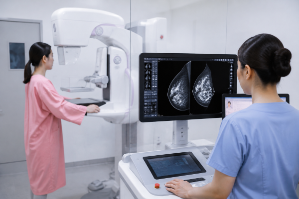
Technical Challenges and Chest Wall Positioning Modifications
Imaging the male chest presents unique physical challenges for technologists. Men possess very little glandular breast tissue. They also have prominent pectoralis major muscles and firm chest walls.
Standard compression plates struggle with flat surfaces. Rigid plates often hit the ribs and the clavicle prematurely. This poor fit causes tissue slippage and introduces motion blur. Radiologists need alternative solutions for these male anatomical obstacles.
Technologists overcome these physical challenges through specialized chest wall positioning adjustments:
- Technologists exchange standard plates for smaller 18×24 cm contact paddles. These smaller paddles compress small tissue volumes directly against the chest wall.
- Technologists apply spot-compression paddles over palpable nodules. These targeted paddles separate overlapping tissue and improve lesion conspicuity.
- Technologists pull mobile lateral tissue away from ribs during chest wall positioning. They rotate the machine gantry to match the angle of the pectoral muscle fibers.
- Technologists use a gentle pinch-and-pull method for the craniocaudal view. This maneuver slides tight subareolar tissue away from underlying muscle bands.
- Modern digital mammography systems automatically adjust tube current. These automated exposure settings penetrate dense tissue without creating beam artifacts.
Proper chest wall positioning ensures complete tissue capture. Technologists image the entire breast tissue volume from the skin to the deep pectoral fascia.
Multimodal Diagnostic Approach Combining Digital Mammography and Ultrasound
Clinicians achieve optimal diagnostic results by pairing digital mammography with high-frequency ultrasound. This multimodal strategy provides comprehensive diagnostic information for complex male cases.
- The clinical workup begins when a patient discovers a suspicious breast mass. Technologists perform diagnostic breast examinations using specialized views.
- Radiologists review the resulting images to evaluate tissue architecture. They assign an initial BIRADS category to record their level of suspicion.
- Technologists perform a targeted high-frequency ultrasound examination immediately after primary x-ray imaging. Ultrasound determines whether the detected mass is solid or fluid-filled.
- Technologists also scan the adjacent axillary regions. This sonographic step evaluates the patient for enlarged or suspicious lymph nodes.
- Physicians perform a core needle biopsy when imaging reveals suspicious features. They use radiographic guidance or ultrasound to direct the biopsy needle.
- Pathologists examine the harvested tissue samples under a microscope. They determine the histological grade and measure hormone receptor levels.
Targeted ultrasound complements digital mammography effectively. However, ultrasound cannot display broad tissue architecture or spot tiny microcalcifications across wide fields. Full-field radiographic views provide the structural roadmap. Doctors need both modalities to confirm disease extent. This combined approach ensures accurate tumor sampling and optimal staging.
Conclusion and Clinical Recommendations for Male Breast Screening
Digital mammography reliably overcomes male anatomical challenges to deliver vital diagnostic clarity. It accurately separates gynecomastia from male breast cancer (MBC). It also highlights dangerous microcalcifications and guides clinical biopsies. This effective tool does not belong solely to female medicine.

