
How to Prevent Common Image Artifacts in Digital Mammography
Preventing image artifacts in digital mammography requires clear controls across patient positioning, detector stability, and exposure settings. Subtle architectural distortions and microcalcifications measuring 100 to 200 micrometers rely entirely on high-frequency contrast. Even minor technical anomalies can easily hide true malignancies or mimic suspicious masses. Radiology teams must stop these visual errors before they compromise clinical diagnostic confidence. This guide outlines practical techniques to ensure clean, diagnostic-quality images on every single scan.
Patient-Related Artifacts: Preventing Motion Blur Through Patient Positioning in Digital Mammography
Patient movement and skin contaminants cause the majority of repeated exposures in routine digital mammography screening. Technologists can resolve most of these issues by pairing good communication with gentle, firm physical control.
Stopping Motion Blur in Digital Mammography with Controlled Compression
Motion blur destroys fine image detail and turns sharp microcalcifications into fuzzy, unreadable smudges. Preventing motion blur in digital mammography demands adequate breast tissue immobilization.
- Target Compression Force: Technologists should apply steady motorized compression between 110 and 150 Newtons.
- Tissue Separation: Proper compression flattens overlapping glandular structures and lowers the required radiation dose.
- Ergonomic Paddles: Flexible paddles distribute pressure evenly across sensitive tissue, minimizing patient flinching.
Clear breathing guidance is equally essential to stop subtle patient flinching during exposure. Technologists should ask the patient to exhale halfway and hold still comfortably for two seconds. This simple breathing cue eliminates sudden chest twitches and stops motion blur entirely.
Optimizing Patient Positioning to Prevent Foreign Residues in Digital Mammography
Everyday cosmetics frequently trick image interpretation software by mimicking dangerous calcification clusters. Common deodorants, antiperspirants, body lotions, and powders contain dense zinc, aluminum, or talc particles. These mineral particles block x-rays and create bright white specks on the final image.
Clinics must provide warm cleansing wipes before every examination to clean the skin properly. Staff should ensure patients wipe their underarms, lower breasts, and collarbones thoroughly.
Careless patient positioning also causes thick skin folds that cast dark shadows across glandular tissue. Technologists should gently smooth loose skin outward toward the chest wall during patient positioning. Keeping the nipple in profile prevents dense tissue overlap from masking true retroareolar lesions.
Hardware Reliability: Flat Panel Detector Artifacts and Ghosting Artifacts in Digital Mammography
Hardware flaws in detector units introduce deceptive linear marks, pixel dropouts, and structural shadows. Both direct conversion and scintillator panels require routine maintenance to keep them operating cleanly according to guidelines from the American College of Radiology
.
Eliminating Ghosting Artifacts and Flat Panel Detector Artifacts
Ghosting artifacts happen when detector materials retain residual electrical charge from a previous high-dose exposure. Stereotactic biopsy procedures and thick calibration blocks frequently cause this stubborn signal lag. The subsequent patient image will show a faint, confusing phantom shadow from the earlier scan.
- Recovery Intervals: Clinics can stop ghosting artifacts by enforcing a thirty-second rest delay between heavy acquisitions.
- Charge Flushing: Technologists must verify that automated optical charge-flushing cycles run fully after each exposure.
- Bad-Pixel Mapping: Staff must run regular flat-field calibrations to fix flat panel detector artifacts through automated pixel mapping.
- Gain Normalization: Regular gain and offset calibrations ensure that every pixel responds with uniform sensitivity across the active field.
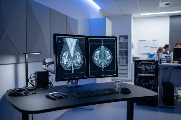
Maintaining Anti-Scatter Grids to Prevent Flat Panel Detector Artifacts in Digital Mammography
Moving anti-scatter grids protect image contrast by absorbing scattered radiation before it hits the detector. Mechanical wear and timing slips in the grid drive quickly ruin image uniformity.
If the oscillating bucky carriage hesitates during exposure, stationary lead lines cast harsh stripes. These fine line shadows spoil image clarity and can mimic dense architectural distortions. Technologists should routinely verify that grid reciprocation matches the tube exposure pulse.
Misalignment between the x-ray tube focal spot and the grid causes severe one-sided grid cut-off. Scheduled engineering checks prevent mechanical drift and keep flat panel detector artifacts away from images.
Software Optimization: Advanced AEC Calibration in Digital Mammography
Incorrect software profiles and poor sensor choices create noisy, washed-out, or overly processed images. Modern screening systems rely on balanced automated exposure algorithms to balance radiation dose and image clarity under
.
Correcting Exposure Errors Through AEC Calibration in Digital Mammography
The Automatic Exposure Control manages tube current and target filtration based on breast tissue attenuation. Serious image errors occur when the active AEC sensor measures the wrong anatomical area.
- Fatty Tissue Cut-Off: If the sensor covers fatty tissue, the machine terminates x-ray exposure prematurely. The resulting underexposure creates quantum mottle that easily conceals subtle architectural distortions.
- Dense Object Overexposure: Placing the sensor over surgical clips or dense implants causes severe overexposure that washes out soft margins.
- Phantom Verification: Medical physicists must perform regular AEC calibration across different acrylic phantom thicknesses.
Modern systems with dynamic multi-cell sensing automatically target the densest tissue regions for optimal exposure.
Balancing Edge Enhancement and AEC Calibration in Digital Mammography
Spatial sharpening filters highlight subtle calcifications, but overly aggressive algorithms introduce deceptive visual artifacts. Heavy edge enhancement often produces a dark, artificial halo around high-density masses and surgical clips.
Diagnostic Warning: The Halo Effect
Excessive high-pass frequency filtration generates an artificial radiolucent line along tissue margins. This fake boundary masks subtle malignant spiculation and distorts microcalcification clusters.
Radiologists and application specialists should work together to tune image processing algorithms carefully. Clinical teams should adjust contrast lookup tables using standard contrast-detail test phantoms. Proper software tuning and verified AEC calibration preserve realistic anatomy on diagnostic review monitors. Balanced post-processing guarantees that radiologists review authentic anatomical details rather than software artifacts.
Key Clinical Takeaways: Advancing Digital Mammography Through Precise Patient Positioning
Preventing image artifacts in digital mammography relies on steady daily habits rather than emergency troubleshooting. Disciplined patient positioning and clear breathing guidance stop motion blur before the exposure starts. Scheduled detector calibration and sensor checks protect your equipment from drift and exposure errors. Every clean scan avoids unnecessary patient radiation and keeps your screening schedule running on time.
Contact our clinical imaging team today to learn how our digital mammography equipment delivers dependable, artifact-free diagnostics.

