
Can a Digital Mammography Machine Safely Examine Patients with Breast Implants?
A digital mammography machine can safely evaluate individuals with silicone or saline breast implants. Clinicians apply validated displacement maneuvers to conduct standard screenings without risking implant rupture.
Millions of women worldwide undergo cosmetic or reconstructive breast augmentation every year. However, augmented individuals frequently worry about procedural safety during annual checkups. Patients often fear mechanical ruptures from direct compression paddles. Furthermore, dense prosthetic shells naturally attenuate primary x-ray photons. This physical barrier blocks underlying parenchymal details on standard radiographic displays.
Fortunately, modern digital mammography systems incorporate advanced pressure sensors to protect fragile capsules. Skilled radiologic technologists also utilize specialized positioning protocols for augmented tissue. Consequently, digital mammography provides dependable early cancer detection while safeguarding structural prosthesis integrity.
Biomechanical Safety Standards and Controlled Clinical Compression Limits
Modern silicone and saline prostheses exhibit extraordinary mechanical resilience against external forces. Industrial manufacturing standards mandate extensive stress testing before regulatory commercial release. Implant shells routinely withstand compressive loads exceeding several hundred pounds of pressure.
In contrast, clinical digital mammography systems apply strictly regulated, gentle compression forces. Technologists apply pressure solely to immobilize tissue and prevent motion blur. The applied load remains far below industrial elastomeric burst limits.
| Evaluation Parameter | Industrial Testing Standard | Clinical Mammography Limit |
| Applied Force | Up to 300 kg static load | 11 to 20 daN controlled load |
| Material Resistance | High-tensile cross-linked silicone | Flexible biocompatible shell |
| Mechanical Goal | Rupture point identification | Motion blur elimination |
| Primary Safety Margin | Extreme physical overload buffer | Multi-sensor feedback shutdown |
Advanced digital mammography units incorporate intelligent microprocessor controls. These motorized paddles monitor resistance continuously across tissue surfaces. Automated feedback sensors prevent localized over-pressurization near firm edges.
The machine automatically halts paddle advancement once optimal tissue tension occurs. This calibrated feedback eliminates sudden pressure spikes against retromammary prostheses. Therefore, modern digital mammography delivers reliable physical safety during every clinical screening.
The Eklund Technique and Specialized Implant Displacement Views
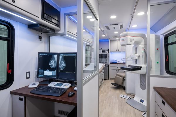
Standard screening protocols require modification to image augmented patients thoroughly. In 1988, Dr. G. W. Eklund designed modified displacement projections. These projections remain the clinical gold standard in modern radiology clinics. Technologists routinely capture eight distinct views instead of standard four-view examinations.
The standard examination begins with light compression projections of both breasts:
- Technologists capture standard craniocaudal projections showing the implant and surrounding margins.
- Technologists obtain mediolateral oblique views to evaluate retromammary regions and axillary tails.
- The imaging platform uses minimal compression to prevent capsular strain during primary views.
- These preliminary exposures document overall implant location, contour, and capsular calcification status.
After capturing initial images, the technologist transitions to specialized implant-displaced views:
- Technologists perform manual palpation to locate anterior implant margins accurately.
- Technologists gently push the prosthesis backward against the rigid chest wall.
- Technologists pull native anterior glandular tissue forward onto the digital detector.
- The compression paddle secures native parenchyma while excluding the dense prosthesis.
These implant-displaced views maximize clear visualization of anterior glandular tissues. Modern digital mammography captures peripheral tissue without obscuring critical retroareolar details. Consequently, digital mammography allows clinicians to detect subtle architectural distortions reliably.
Exposure Parameter Adjustments and Diagnostic Radiographic Boundaries
Prosthetic materials possess high radiodensity compared to surrounding human soft tissues. Saline and silicone attenuate x-ray photons significantly more than glandular structures. This density differential challenges standard automated exposure control systems.
Standard automatic sensors measure primary photon attenuation across broad detector areas. When an opaque implant covers sensor cells, systems increase radiation output substantially. This automated increase causes severe overexposure along thin peripheral skin boundaries.
Technologists bypass this imaging error through manual parameter management:
- Technologists deactivate standard automated exposure sensors during implant-displaced views.
- Technologists select manual tube potential settings to balance penetration depth and contrast.
- Operators choose specialized target-filter combinations to optimize low-dose energy spectra.
- Modern digital mammography software applies dynamic multi-frequency algorithms to equalize peripheral margins.
Radiologists must also identify pre-existing implant complications prior to mechanical compression. Severe fibrous capsular contracture hardens breast tissue, making compression painful and difficult.
Furthermore, silent intracapsular ruptures can develop spontaneously over prolonged implantation periods. Technologists conduct thorough physical examinations and clinical history reviews beforehand. When clinicians suspect acute ruptures, they refer patients for diagnostic breast MRI. Nevertheless, routine screening via digital mammography remains the frontline baseline for stable patients.
Clinical Value of Routine Digital Mammography for Augmented Patients
Early detection of breast malignancies remains crucial for all women regardless of augmentation status. Breast implants do not increase malignancy risks, yet they present diagnostic challenges. Regular screening with digital mammography identifies early-stage invasive carcinomas and microcalcifications effectively.
The advanced image processing capabilities of digital mammography support high diagnostic confidence:
- Flat-panel digital detectors provide high spatial resolution for microcalcification detection.
- Advanced software sharpens contrast between glandular tissues and dense silicone borders.
- Radiologists review digital displays instantly, reducing recall rates for additional views.
- Clinics transfer electronic files rapidly to support multidisciplinary oncology consultations.
Consistent imaging histories enable radiologists to detect tiny interval architectural modifications. Modern digital mammography platforms empower clinics to deliver compassionate, safe care. Patients maintain their cosmetic results while protecting their long-term health.
Upgrade Your Clinic With Implant-Safe Digital Mammography Platforms
Validated displacement techniques ensure completely safe breast cancer screening for augmented patients. Advanced digital mammography hardware prevents mechanical trauma while delivering superior diagnostic image clarity. Modern healthcare facilities need reliable imaging tools to accommodate diverse patient populations.
Are you looking to modernize your diagnostic radiology department? Our cutting-edge digital mammography systems feature intuitive displacement software and smart pressure management. Contact our technical team today to request product specifications, clinical demonstrations, and competitive procurement quotes.

