
How Does Dynamic Digital Radiography Work?
Dynamic digital radiography works by firing ultra-short, pulsed X-ray bursts synchronized with a high-speed flat-panel detector. This system acquires rapid sequential image series that reveal internal structural movement at remarkably low radiation doses.
Traditional radiographic examinations provide exceptional spatial resolution for bone and soft tissue structures. However, conventional projection radiography captures only frozen, static representations of anatomical systems. Human anatomy relies on active kinetic coordination across complex cardiopulmonary, musculoskeletal, and vascular structures. Static images frequently miss dynamic joint impingement, intermittent airway narrowing, or asymmetrical diaphragmatic movement.
Fluoroscopy provides real-time movement analysis, but standard systems deliver lower spatial resolution alongside substantial cumulative radiation burdens. To bridge this critical diagnostic gap, medical engineering introduced dynamic imaging. This innovative modality merges the crisp clarity of static radiography with continuous functional motion capture. Clinicians now evaluate living biomechanics in upright, weight-bearing postures without subjecting patients to excessive radiation levels.
Hardware Architecture and Pulse Synchronization in Dynamic Digital Radiography
The diagnostic performance of dynamic digital radiography depends directly on specialized generator control and detector synchronization. Conventional continuous fluoroscopy exposes patients to an uninterrupted beam of ionizing radiation throughout the examination period. In contrast, pulsed X-ray imaging employs millisecond pulse modulation to capture individual high-resolution projections.
| System Parameter | Conventional Fluoroscopy | Dynamic Digital Radiography (DDR) |
| X-Ray Beam Profile | Continuous exposure beam | Ultra-short pulsed exposure |
| Pulse Duration per Frame | Continuous or 10 to 30 ms | 1 to 5 ms ultra-short pulse |
| Detector Frame Rate | Standard video rates | 15 to 30 frames per second |
| Total Diagnostic Dose | Moderate to high accumulation | Comparable to two-view static X-ray |
| Clinical Posture | Typically recumbent fluoroscopy | Natural upright, weight-bearing |
Modern dynamic digital radiography platforms combine three tightly integrated hardware subsystems to guarantee clinical safety and diagnostic precision:
- A high-frequency generator fires ultra-short X-ray pulses with exposure times lasting only a few milliseconds per frame.
- An ultra-fast flat-panel detector collects transmitted photons instantly, capturing between 15 and 30 discrete frames per second.
- Microprocessor synchronization circuitry aligns each distinct radiation burst perfectly with active detector read cycles.
- Real-time automated dose modulation continuously measures patient thickness, adjusting beam intensity instantly across the motion sequence.
By pulsing radiation instead of maintaining a continuous beam, dynamic radiographic prevents motion-induced edge blurring. Patients complete a dynamic sequence lasting several seconds while receiving total radiation comparable to routine static imaging.
High-Speed Flat-Panel Detector Architecture and Direct Conversion
Advanced detector physics underpins the image quality achieved by dynamic digital radiography. Early digital imaging devices lacked the data processing bandwidth required to capture successive full-resolution radiographs simultaneously. Modern serial digital imaging utilizes high-speed flat-panel detectors built with rapid direct-deposition scintillation layers and active matrix backplanes.
- High-purity cesium iodide scintillators convert incoming pulsed X-ray photons into visible light waves with minimal lateral dispersion.
- Amorphous silicon thin-film transistor arrays capture optical signals rapidly, converting light photons directly into digital electrical charges.
- Low-noise readout electronics process multi-channel pixel data in parallel, supporting acquisition rates of up to 30 frames per second.
- Ghost-suppression engineering prevents image lag by purging residual electrical charges from pixel storage capacitors between consecutive frames.
These architectural advancements ensure that dynamic flat-panel radiography maintains high modulation transfer functions throughout rapid movements. The resulting image sequence features sharp structural margins, high dynamic range, and consistent pixel clarity across every frame.
Image Processing and Optical Flow Analysis Algorithms
Generating clear clinical cinema requires sophisticated post-processing pipelines. A single dynamic digital radiography examination produces hundreds of raw projection frames within a few seconds of patient movement. Without automated image processing, random scatter radiation and quantum noise could obscure faint anatomical boundaries.
Modern dynamic digital radiography systems process acquired image stacks through sequential algorithmic enhancement stages:
- Temporal recursive filtration identifies and suppresses random quantum noise fluctuations across consecutive frames without degrading anatomical edge sharpness.
- Multi-frequency contrast algorithms dynamically equalize bright air-filled lung spaces and dense mediastinal structures across dynamic breathing cycles.
- Scatter-correction software estimates photon dispersal patterns numerically, eliminating the need for bulky physical anti-scatter grids.
- Optical flow analysis algorithms track regional pixel luminosity shifts across successive frames to compute local anatomical velocity fields.
- Spatial cross-correlation algorithms map tissue displacement vectors, highlighting subtle structural discrepancies across adjacent anatomical zones.
Through advanced algorithmic analysis, dynamic digital radiography transforms raw video frames into quantitative maps of physical movement. Radiologists observe dynamic structural interaction with exceptional diagnostic confidence and physiological precision.
Measuring Diaphragmatic Excursion and Joint Kinetics
The primary advantage of dynamic digital radiography lies in its unique ability to quantify mechanical tissue movement. Clinicians no longer rely exclusively on qualitative visual impressions of static radiographic images. Instead, dynamic digital radiography yields numerical velocity measurements, spatial trajectory data, and expansion ratios across active organ systems.
The core diagnostic workflow proceeds systematically through established processing stages:
- The system acquires sequential pulsed projection frames at rates between 15 and 30 frames per second.
- Image processing software applies temporal filtration and optical flow algorithms to map velocity vectors.
- For cardiopulmonary assessment, software calculates bilateral diaphragmatic excursion and maps regional ventilation.
- For musculoskeletal kinematics, algorithms track joint stability, patellar tracking paths, and dynamic ligamentous laxity.
Clinicians leverage dynamic digital radiography across diverse clinical specialties to evaluate complex kinetic conditions:
- Radiologists measure bilateral diaphragmatic excursion directly, quantifying vertical diaphragm displacement in centimeters throughout full respiratory cycles.
- Pulmonologists evaluate regional lung ventilation abnormalities, utilizing pixel densitometry changes to identify air trapping in chronic obstructive pulmonary disease.
- Thoracic surgeons verify diaphragmatic paralysis caused by phrenic nerve trauma by observing paradoxical upward motion during forced inhalation.
- Orthopedic specialists monitor dynamic joint instability under physiological weight-bearing loads, identifying ligamentous laxity and occult subluxations.
- Clinicians evaluate postoperative orthopedic implant mechanics, detecting subtle micro-motion and mechanical loosening during active joint flexion.
By capturing real-time physiological mechanics, dynamic digital radiography uncovers hidden functional pathologies that remain invisible during standard examinations.
Clinical Workflow and Efficiency of Dynamic Digital Radiography
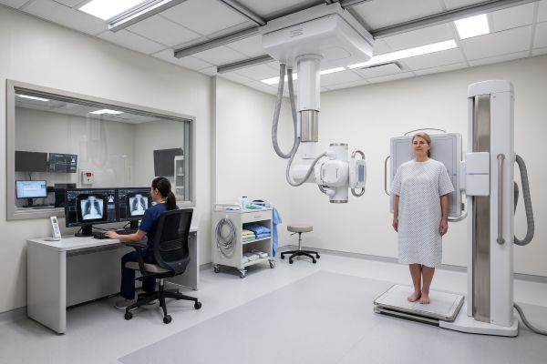
Integrating dynamic digital radiography into clinical operations requires no disruptive alterations to standard radiographic suites. Modern systems utilize multifunctional digital X-ray hardware capable of executing routine static projections alongside advanced dynamic acquisitions.
Healthcare facilities achieve marked operational and economic advantages by deploying dynamic digital radiography platforms:
- Technologists perform static and dynamic examinations within a single diagnostic room, maximizing space efficiency and system utilization rates.
- Patients complete dynamic kinetic evaluations within seconds while standing comfortably in natural, functional weight-bearing postures.
- Diagnostic throughput increases substantially because rapid acquisition protocols eliminate tedious multi-view static positioning adjustments.
- Inter-observer diagnostic agreement improves because objective mathematical vectors replace subjective visual assessments of joint motion.
- Healthcare institutions expand clinical offerings, attracting referrals from sports medicine, orthopedic surgery, and pulmonary rehabilitation departments.
Consequently, dynamic digital radiography represents a practical, high-value modernization pathway for progressive imaging centers seeking advanced functional capabilities.
Elevate Your Diagnostic Imaging With Dynamic Digital Radiography
Dynamic digital radiography bridges the historical divide between high-resolution static radiography and real-time fluoroscopic motion assessment. By firing synchronized, ultra-short pulses through high-speed flat-panel detectors, this modality visualizes anatomical movement at minimal radiation levels. Advanced motion-tracking software translates complex biomechanics into reproducible diagnostic metrics, empowering physicians to diagnose disorders earlier and more accurately.
Are you prepared to expand your department’s clinical diagnostic reach? Our cutting-edge dynamic digital radiography systems integrate easily into existing hospital environments, delivering exceptional image quality, intuitive workflow software, and comprehensive patient protection. Contact our clinical technical team today to schedule an equipment demonstration, discuss technical specifications, and receive a customized facility quotation.

