Can Dynamic Digital Radiography Be Used for Cardiac Imaging?
Dynamic digital radiography can be used for cardiac imaging in specific clinical and research applications to observe heart kinetics, cardiac silhouette deformation, and thoracic interaction in real time. Because cardiac contractions are inherently rapid and cyclical, conventional static radiographs only capture a random, instantaneous frame within the continuous heartbeat. Dynamic digital radiography addresses this limitation by acquiring continuous low-dose pulsed X-ray frames across multiple cardiac cycles, providing essential temporal motion information that planar projections cannot deliver. However, it functions as a functional radiographic screening and biomechanical evaluation tool rather than a replacement for specialized cross-sectional or volumetric cardiovascular modalities.
Dynamic Digital Radiography Records Cardiac Motion Over Time
Cardiac mechanics involve continuous, coordinated physical contractions that change rapidly with every millisecond of the cardiac cycle. Evaluating these kinetic transitions requires radiographic technology capable of tracking anatomical shifts over time.
Dynamic digital radiography records this continuous movement by acquiring high-frequency pulsed radiographic series. Instead of exposing a single static film, the system deploys a rapid series of pulsed exposures while the patient remains naturally positioned. Sequential multi-frame cine loops capture wall excursion, systolic volume contraction, and diastolic expansion. Introducing this temporal dimension directly into planar projection imaging allows clinicians to inspect cyclic border displacement that would remain invisible on isolated still radiographs.
Dynamic Imaging Shows Cardiac and Thoracic Interactions
Beyond tracking the heart wall in isolation, dynamic digital radiography provides a comprehensive panoramic view of thoracic kinetics, revealing how cardiac motion interacts with surrounding respiratory mechanics.
During thoracic dynamic acquisitions, clinicians observe synchronized heart silhouette shifts alongside diaphragmatic movement and lung expansion. This real-time perspective helps medical professionals evaluate pericardial adherence, verify cardiac pulsation transmission to neighboring pulmonary tissue, and assess structural shifts during deep inhalation or forced breath-holding. While conventional static chest radiographs establish anatomical baselines, the dynamic cine series shows the physical interplay between cardiovascular structures and thoracic breathing biomechanics.
| Imaging Modality | Primary Diagnostic Output | Imaging Perspective | Primary Clinical Focus |
| Dynamic Digital Radiography | Low-dose planar dynamic cine series | Wide-field upright projection | Functional motion tracking and thoracic kinetic interaction |
| Echocardiography | Real-time acoustic Doppler and tissue tracking | Focused sectional acoustic windows | Valvular morphology, intracardiac hemodynamics, and ejection fraction |
| Computed Tomography | High-resolution isotropic 3D volumetric slices | Multiplanar tomographic reconstructions | Coronary calcium scoring and lumen stenosis quantification |
| Magnetic Resonance Imaging | Multiparametric tissue and velocity sequences | Slice-selective cross-sectional views | Myocardial viability, tissue characterization, and flow dynamics |
Image Analysis Adds Quantitative Functional Information
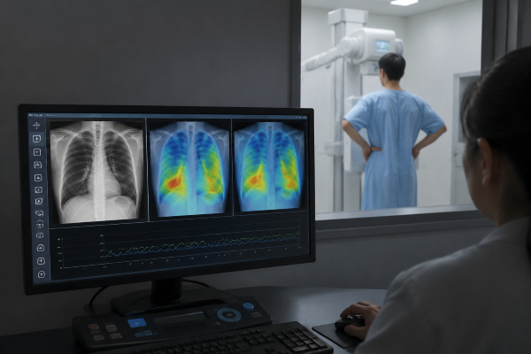
Modern post-processing platforms turn raw dynamic X-ray loops into objective quantitative kinetic parameters. These analytical pipelines convert observed spatial movement into reproducible diagnostic data.
Specialized analytical software extracts structural displacement metrics, tracing border contraction velocity, regional motion amplitude, and dynamic waveform curves from sequential projection frames. Correlating regional pixel intensity variations with cardiac pulsation cycles allows medical researchers to visualize localized motility patterns. The breadth and precision of these quantitative assessments depend on installed software packages and detector performance parameters.
Dynamic Digital Radiography Complements Established Modalities
Dynamic digital radiography serves as a broad functional radiographic tool rather than a replacement for dedicated cardiovascular imaging modalities. Comprehensive clinical workups rely on distinct diagnostic strengths tailored to specific pathologies.
Echocardiography delivers non-ionizing, detailed valvular hemodynamics, while computed tomography provides exquisite sub-millimeter visualization of coronary arterial anatomy and calcification. Similarly, cardiac magnetic resonance imaging remains the recognized benchmark for tissue characterization and myocardial perfusion. The unique advantage of dynamic digital radiography lies in capturing functional movement within standard wide-field planar X-ray workflows at substantially lower radiation doses and equipment complexity than volumetric modalities.
Conclusion
Dynamic digital radiography can be utilized for targeted cardiac evaluations, capturing cyclic heart wall kinetics, silhouette shifts, and cardiopulmonary biomechanical interactions over time. Its key value lies in adding continuous temporal information to planar X-ray imaging without complicating patient positioning. Final diagnostic capability ultimately depends on specific detector frame rates, software analytical packages, and institutional clinical protocols.
Contact our technical specialists today to discuss clinical imaging workflows and explore dynamic digital radiography solutions for your facility.