
Dynamic Digital Radiography for Respiratory Assessment
Respiratory diseases often affect lung function before obvious structural abnormalities become visible on conventional X-ray images. As a result, evaluating how the lungs and diaphragm move during breathing has become increasingly important in clinical practice. Dynamic Digital Radiography for Respiratory Assessment offers a practical way to combine anatomical imaging with functional evaluation in a single examination. By capturing a sequence of digital X-ray images during natural respiration, DDR provides additional insight into respiratory mechanics. This functional information cannot be obtained from a standard chest radiograph alone.
Dynamic Digital Radiography in Clinical Respiratory Practice
Conventional Digital Radiography remains the first-line imaging method for many respiratory conditions because it is fast, widely available, and cost-effective. However, a static chest radiograph represents only one moment in the breathing cycle. Patients with diaphragm dysfunction or impaired lung expansion may still present with normal or inconclusive radiographic findings. As a result, diagnosis can become more challenging.
Dynamic Digital Radiography addresses this limitation by recording continuous image sequences while the patient breathes normally. The examination goes beyond anatomical structures. It shows how the diaphragm, lungs, and chest wall move throughout the respiratory cycle.
This additional functional information helps clinicians better understand respiratory abnormalities and supports more confident clinical decision-making.
Clinical Applications of Dynamic Digital Radiography for Respiratory Assessment
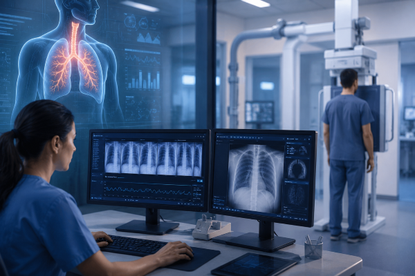
Growing clinical evidence has demonstrated the value of Dynamic Digital Radiography in respiratory medicine. One well-documented application is the assessment of diaphragm dysfunction, where dynamic motion plays a key role in diagnosis.
A published case report described a 46-year-old man with unexplained dyspnea and reduced exercise tolerance. Although a conventional chest radiograph showed an elevated right hemidiaphragm, the underlying cause remained uncertain. Dynamic Digital Radiography was subsequently performed while the patient breathed normally. The examination clearly demonstrated reduced movement of the right diaphragm throughout the respiratory cycle. This functional information supported the diagnosis of diaphragmatic dysfunction. This case illustrates how motion analysis can complement conventional imaging when structural findings alone are insufficient.
Researchers have also investigated Dynamic Digital Radiography in patients with chronic obstructive pulmonary disease (COPD). By analyzing changes in lung area, diaphragm excursion, and chest wall motion during breathing, clinicians can better evaluate respiratory mechanics and monitor functional impairment alongside routine imaging findings.
Evaluating Pulmonary Function with DDR
Pulmonary function tests remain an essential part of respiratory assessment, but they provide numerical measurements rather than direct visualization of respiratory motion. Imaging techniques that reveal how the lungs function during breathing can therefore provide valuable complementary information.
Recent studies have shown that Dynamic Digital Radiography can quantify diaphragm excursion, lung area changes, and respiratory motion using dedicated image analysis. These measurements are generated directly from the dynamic image sequence. Clinicians can evaluate anatomical structures and respiratory motion during the same examination. Because the workflow is similar to routine Digital Radiography, the additional information can be obtained without introducing a completely different imaging procedure.
Improving Follow-Up and Long-Term Patient Management
Respiratory assessment does not end once a diagnosis has been made. Patients recovering from thoracic surgery, receiving treatment for chronic lung disease, or participating in pulmonary rehabilitation often require repeated evaluations to monitor functional changes over time.
Because Dynamic Digital Radiography follows a workflow similar to routine Digital Radiography, repeat examinations can be incorporated into regular clinical practice with minimal disruption. Comparing dynamic image sequences from different visits helps clinicians identify changes in diaphragm motion and lung expansion. These findings may support the evaluation of treatment response and functional recovery. Rather than relying solely on symptom reports or static images, physicians can assess how respiratory function evolves throughout the course of care.
The Future of Dynamic Digital Radiography for Respiratory Assessment
Research into Dynamic Digital Radiography for Respiratory Assessment continues to expand. Recent studies are exploring automated image analysis techniques for dynamic image sequences. These methods can measure diaphragm excursion, lung area variation, and respiratory motion more efficiently. These quantitative measurements may improve consistency between examinations while reducing the time required for image interpretation.
As artificial intelligence becomes increasingly integrated into medical imaging, future DDR systems are expected to provide more advanced functional analysis and clinical decision support. Although the technology is still evolving, current evidence suggests that dynamic respiratory imaging will become an increasingly valuable complement to conventional chest radiography.
Conclusion
Accurate respiratory assessment requires more than identifying structural abnormalities. Understanding how the lungs and diaphragm move throughout the breathing cycle can provide important functional information that supports diagnosis, treatment planning, and long-term patient management.
DDR combines real-time motion analysis with familiar Digital Radiography workflows. This approach enables respiratory function to be evaluated without significantly changing routine imaging procedures. As clinical evidence continues to grow, the technology is expected to play an increasingly important role in modern respiratory imaging.
At SEEHO Medical Equipment, we are committed to delivering advanced Digital Radiography solutions that meet the evolving needs of modern hAs Dynamic Digital Radiography continues to gain recognition in respiratory imaging, healthcare providers are looking for solutions that combine reliable performance with efficient clinical workflows. SEEHO Medical Equipment provides advanced Dynamic DR systems designed to support modern diagnostic practice. We work closely with hospitals and healthcare institutions to deliver imaging solutions that align with their clinical requirements and long-term development goals.ealthcare. If you would like to learn more about our Dynamic DR systems or discuss the best imaging solution for your clinical practice, our experienced team is ready to provide professional support and tailored recommendations.

