
How Does Dynamic Digital Radiography Support Functional Imaging?
Dynamic digital radiography supports functional imaging by introducing the dimension of time to planar X-ray acquisition, capturing physiological motion rather than isolated anatomy. While conventional static radiographs record high-contrast morphological snapshots at a single moment, functional imaging requires evaluating how anatomical structures behave during active movement. Dynamic DR bridges this gap by acquiring sequential radiographic frames at low exposure levels across physiological cycles. Consequently, clinicians can observe continuous biomechanical excursion and respiratory mechanics that remain invisible on static examinations.
Dynamic Imaging Shows Changes During Physiological Motion
Continuous image acquisition captures structural displacement throughout active physiological motion. In thoracic examinations, respiration involves complex interactions between the diaphragm, thoracic cage, and pulmonary parenchyma. A standard static radiograph reveals lung volume only at full inspiration or expiration, leaving intermediate mechanics unrecorded.
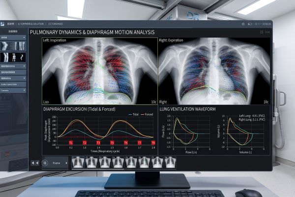
Dynamic digital radiography records the entire respiratory cycle from inspiration through exhalation. This dynamic sequence visualizes diaphragmatic excursion, chest wall expansion, and regional airway caliber changes in real time. Comparing sequential dynamic frames to an isolated static image highlights distinct diagnostic layers: static views verify anatomical presence, whereas dynamic sequences illustrate physical performance and functional continuity.
Dynamic Digital Radiography Can Support Functional Assessment
Dynamic radiographic sequences provide valuable clinical visual data that assist medical teams in functional diagnosis. In pulmonary medicine, dynamic digital radiography can support the evaluation of conditions characterized by impaired ventilation dynamics, such as chronic obstructive pulmonary disease (COPD) or diaphragm paralysis. Clinicians can observe paradoxical motion, delayed regional ventilation, or restricted excursion during continuous breathing maneuvers.
Beyond thoracic applications, the technology assists orthopedic specialists in evaluating musculoskeletal kinematics. When patients flex, extend, or rotate joints under natural physical loading, dynamic dr helps clinicians track joint congruency, patellar tracking, and subtle instability. Dynamic visualization supports clinical judgment by demonstrating motion patterns, though definitive clinical diagnoses rely on comprehensive patient evaluation alongside complementary testing.
| Diagnostic Focus | Static Radiographic Capability | Dynamic DR Functional Support |
| Thoracic Evaluation | Static lung volume and baseline parenchymal opacity | Continuous diaphragmatic kinetics and respiratory excursion |
| Musculoskeletal Joints | Bone morphology, alignment, and joint space narrowing | Dynamic joint tracking, ligament stability, and kinematics under load |
| Clinical Value | Anatomical baseline and structural screening | Movement-based physiological insight and functional context |
Image Analysis Turns Motion Into Measurable Information
The time-resolved image stacks acquired during dynamic digital radiography establish the foundation for objective motion analysis. Tracking specific anatomical points across successive frames allows software tools to evaluate trajectory displacement, structural velocity, and overall range of motion.
These functional sequences transform qualitative visual motion into structured datasets for clinical review. Medical software can map boundary displacement over time, helping clinicians correlate visible movement with physiological symptoms. While deeper post-processing models handle complex numerical extractions, dynamic acquisition itself provides the indispensable time-series data required for all downstream analytical evaluation.
Conclusion
Dynamic digital radiography supports functional imaging by combining radiographic clarity with temporal motion tracking. Adding the time dimension does not replace conventional static projection; rather, it supplements anatomical baselines with vital functional context. By visualizing real-time physiological movement, dynamic digital radiography equips clinical teams with comprehensive data to evaluate complex thoracic and musculoskeletal dynamics.
Ready to enhance your functional diagnostic capabilities? Contact our technical team today to request specifications or discuss clinical imaging solutions for your facility.

