
How Modern Digital Mammography Optimizes Early Microcalcification Detection
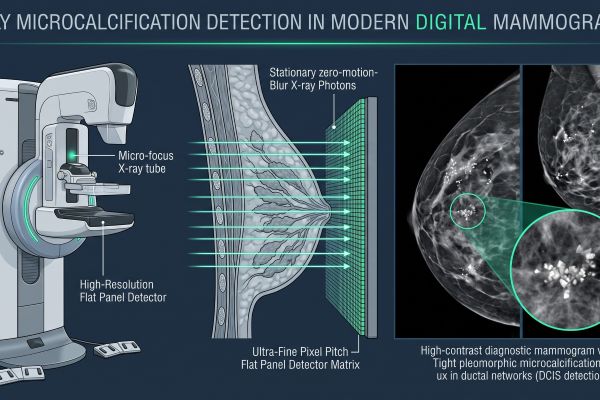
Modern digital mammography optimizes early microcalcification detection by combining ultra-high-pixel-density flat panel detectors with stationary, zero-motion-blur X-ray exposure. Because clustered pleomorphic microcalcifications represent the earliest radiologic hallmark of ductal carcinoma in situ (DCIS), identifying these sub-millimeter mineral deposits before a palpable mass forms is critical for successful clinical intervention. Modern full-field systems maximize diagnostic yield through optimized detector physics, stable micro-focus X-ray generation, and targeted image post-processing algorithms.
Why Microcalcifications Are Crucial in Early Breast Diagnosis
Microcalcifications are tiny calcium deposits within breast tissue that range from 100 μm to several hundred micrometers in diameter. Their radiological characteristics provide essential diagnostic clues regarding cellular activity.
Morphology and Clustering Patterns of Suspicious Calcifications
Evaluating microcalcifications requires assessing both structural morphology and distribution within the breast parenchyma:
- Suspicious Morphologies: Benign calcifications are typically round, rim-like, or coarse. Malignant calcifications linked to DCIS present as fine pleomorphic, linear, or branching forms that trace the inner contours of necrotic ductal lumens.
- Clustering Criteria: A cluster is conventionally defined as five or more microcalcifications confined within a 1 cm³ volume of tissue. Detecting subtle differences in particle shape across these tight clusters demands high structural fidelity from the imaging chain.
Overcoming Tissue Background Noise with High Pixel Pitch
Dense fibroglandular tissue creates anatomic noise that obscures fine structural details. Conventional radiography often suffers from contrast degradation in radiographically dense breasts. Modern digital mammography overcomes this masking effect by using fine pixel pitches (50 μm to 85 μm) and specialized selenium- or cesium-based conversion layers. These technical parameters suppress electronic noise, optimize the contrast-to-noise ratio (CNR), and prevent microcalcifications from blending into dense parenchymal backgrounds.
Technical Strengths of Modern Digital Mammography Systems
Hardware engineering in modern digital mammography ensures maximum spatial sharpness and structural stability during every exposure.
| Hardware Subsystem | Technical Implementation | Diagnostic Impact on Microcalcifications |
|---|---|---|
| Flat-Panel Detector (FPD) | Direct conversion (a-Se) or refined columnar CsI:Tl | Maximizes high-frequency Modulation Transfer Function (MTF) to preserve crisp margins |
| X-Ray Tube Focal Spot | Dual-focus (0.1 mm micro-focus / 0.3 mm standard focus) | Minimizes geometric penumbra during contact and magnification exposures |
| High-Frequency Generator | 50–100 kHz high-voltage output with precise mAs control | Delivers stable X-ray spectra with minimal voltage ripple to reduce motion artifacts |
| Gantry Mechanics | Rigid motorized C-arm with zero mechanical vibration | Eliminates system shake during high-resolution stationary acquisition |
Stationary High-Resolution Flat-Panel Image Acquisition
While multi-angle scanning modalities offer volumetric depth, stationary 2D digital mammography remains unmatched in native in-plane spatial resolution. By maintaining a fixed geometric relationship between the micro-focus tube and the flat panel detector, the acquisition chain completely eliminates the focal spot motion blur associated with sweep mechanisms. This stationary configuration delivers the high spatial frequencies needed to visualize microcalcification margins without interpolation artifacts.
High-Frequency Generators and Stable Micro-Focus X-Ray Tubes
Achieving high resolution requires minimizing geometric unsharpness:
- Micro-Focus Spot Sizes: Using a dedicated 0.1 mm small focal spot for geometric magnification reduces penumbra effects, yielding sharp edge demarcation.
- Target/Filter Combinations: Automated tube design pairs molybdenum (Mo), rhodium (Rh), or tungsten (W) targets with optimized K-edge filters (such as Rh or Ag). This configuration produces a tailored low-energy X-ray spectrum (25–35 kVp) that accentuates photoelectric absorption in calcium deposits while controlling radiation dose.
Advanced Image Post-Processing in Digital Mammography
Hardware acquisition is paired with real-time digital image processing engines to highlight subtle microcalcification clusters without introducing artificial noise.
Dynamic Range Optimization and Contrast Enhancement
Raw digital images possess wide dynamic range but low native contrast across soft-tissue boundaries. Advanced image processing engines apply multi-scale frequency decomposition to separate structural data from low-frequency background gradients:
- Contrast Equalization: Local contrast algorithms amplify high-frequency signals corresponding to mineral grains without overexposing adjacent skin lines or underexposing dense fibroglandular centers.
- Noise Suppression Filters: Adaptive spatial filters smooth random quantum noise while preserving sharp boundaries, ensuring clear visualization of calcification margins.
Magnification Views and Spot Compression Tools
For equivocal findings, digital mammography systems deploy specialized physical and software tools:
- Spot Compression Paddles: Applying localized pressure displaces overlying parenchymal tissue, flattening overlapping structures to isolate suspicious clusters.
- Geometric Magnification (1.5x to 2.0x): Raising the breast support platform above the detector increases optical enlargement on the detector matrix, while software magnification tools provide pixel-level interpolation without degrading diagnostic contrast.
Strategic Summary
High-resolution 2D digital mammography remains the gold standard for early microcalcification detection and DCIS screening. By combining fine-pitch flat panel detectors, vibration-free stationary acquisition, stable micro-focus X-ray tubes, and specialized contrast processing, modern systems deliver the diagnostic confidence required to detect early-stage breast lesions.
Contact our medical imaging specialists to review high-definition system specifications, evaluate dynamic phantom imaging data, and configure an advanced digital mammography solution tailored to your clinical workflow.

