
How to Choose the Right Flat Panel Detector for Digital Mammography: a-Se vs. a-Si Explained
Selecting the right flat panel detector for digital mammography comes down to balancing spatial resolution against environmental resilience and cost: choose amorphous Selenium (a-Se) for maximum microcalcification detail, or amorphous Silicon (a-Si) paired with Cesium Iodide (CsI) for thermal stability, mechanical durability, and long-term economic efficiency. While a-Se relies on direct conversion to prevent optical blur, indirect a-Si detectors leverage high light-yield scintillators to reduce radiation requirements. Evaluating detector architecture, imaging physics, and clinical throughput ensures your facility invests in the optimal diagnostic solution.
Direct vs. Indirect Conversion in Digital Mammography
Flat panel detectors (FPDs) in digital mammography are broadly categorized by how they convert incident X-ray photons into measurable electrical charge.
How Direct Conversion (a-Se) Captures Sharp X-Ray Photons
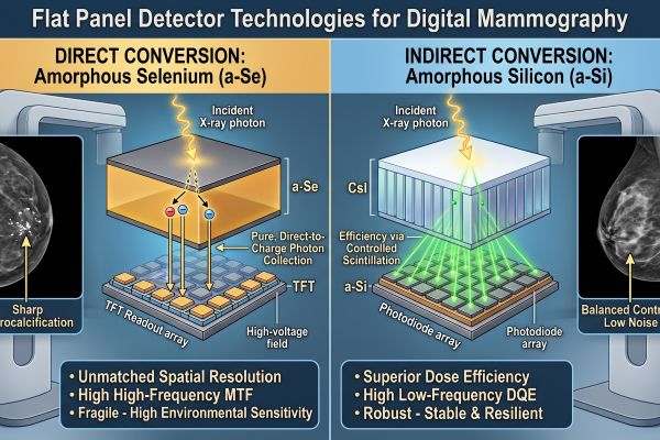
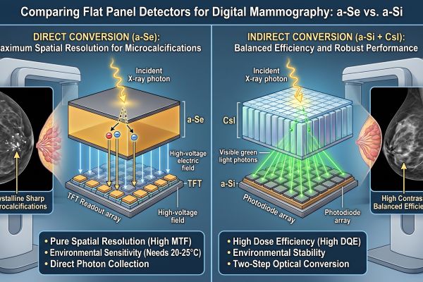
Direct conversion detectors use a layer of amorphous selenium (a-Se) biased with a high-voltage electrical field. When X-ray photons strike the a-Se layer, electron-hole pairs generate directly within the photoconductor:
- Direct Charge Collection: An applied electric field pulls liberated charges straight down to the thin-film transistor (TFT) readout array with virtually zero lateral dispersion.
- Preservation of Fine Details: Because no intermediate optical conversion takes place, direct conversion avoids light scattering. This delivers crisp boundary delineation for minute anatomical structures such as tiny pleomorphic microcalcifications.
How Indirect Conversion (a-Si + CsI) Balances Efficiency and Durability
Indirect conversion detectors utilize a structured scintillator layer—typically thallium-doped Cesium Iodide (CsI:Tl)—bonded to a hydrogenated amorphous silicon (a-Si) photodiode array.
- Two-Step Conversion: Incident X-rays hit the CsI needle crystal structure, which converts photon energy into visible green light. The a-Si photodiode array beneath then converts that light into an electronic signal.
- Optical Waveguiding: Columnar needle structures in modern CsI channel light downward toward the pixels to limit lateral spread, achieving a balance between high quantum efficiency and structural robustness.
Technical Comparison: Resolution, Dose Efficiency, and Stability
Choosing between a-Se and a-Si requires examining three core metrics: spatial resolution, radiation dose efficiency, and environmental operating thresholds.
| Performance Metric | Direct Conversion (a-Se) | Indirect Conversion (a-Si + CsI) |
| Pixel Pitch Range | 50 μm – 85 μm | 70 μm – 100 μm |
| High-Frequency MTF | Exceptional (minimal blur) | Moderate to High (limited by light diffusion) |
| Low-Dose DQE (at low lp/mm) | Moderate | High (superior light yield from CsI) |
| Temperature Tolerance | Strict (requires 20°C–25°C 24/7 cooling) | Broad (resilient to ambient shifts) |
| Mechanical Shock Resistance | Fragile (vulnerable to thermal shock/impact) | High (stable glass/photodiode bonding) |
Spatial Resolution and MTF (Modulation Transfer Function)
Modulation Transfer Function (MTF) reflects how faithfully a detector preserves object contrast at escalating spatial frequencies. In digital mammography, early breast cancer detection depends on resolving microcalcifications as small as 100–200 μm. Because a-Se avoids light diffusion, its MTF remains superior at high spatial frequencies (e.g., >5 lp/mm), yielding sharply defined margins.
DQE (Detective Quantum Efficiency) at Low Radiation Doses
Detective Quantum Efficiency (DQE) evaluates how effectively a detector transfers the signal-to-noise ratio (SNR) from incoming X-rays to the final digital image. At lower spatial frequencies and reduced radiation doses, structured CsI scintillators achieve high absorption efficiency and photon gain. This allows indirect digital mammography systems to produce diagnostic-quality images with low patient radiation exposure.
Environmental Temperature and Mechanical Sensitivity
Operational resilience often separates laboratory performance from real-world reliability:
- a-Se Vulnerabilities: Amorphous selenium has a low glass transition temperature (~35°C–40°C). Exposure to fluctuating temperatures or cooling failures can trigger irreversible crystallization, causing permanent detector artifacts. Continuous 24/7 climate control is mandatory.
- a-Si Durability: Amorphous silicon and CsI assemblies tolerate wider temperature shifts, humidity ranges, and mechanical vibrations, making them dependable across varied clinical environments.
Choosing the Best Detector for Your Digital Mammography Clinic
Aligning detector physics with your facility’s operational workflow prevents costly downtime and diagnostic compromises.
Clinical Workload and High-Throughput Screening Considerations
For dedicated breast imaging centers where diagnostic mammograms, stereotactic biopsies, and complex characterizations dominate, a-Se provides the spatial fidelity required for subtle tissue differentiation.
Conversely, high-throughput screening clinics, mobile mammography units, and regional hospitals benefit significantly from a-Si flat panel detectors. Their rapid frame rates, robust thermal tolerance, and consistent low-dose image acquisition keep patient flow moving without sensitive environmental calibration pauses.
Long-Term Maintenance and Detector Replacement Economics
Total cost of ownership extends beyond initial equipment procurement:
- HVAC and Power Backup: Operating an a-Se digital mammography unit requires uninterruptible power supply (UPS) integration for cooling systems to prevent crystallization during grid outages.
- Replacement Costs: In mobile or semi-controlled clinical settings, a-Si arrays offer longer operational lifespans and lower failure rates due to their mechanical durability.
- Clinical Integration: Modern indirect digital mammography systems deliver sufficient MTF for routine screening protocols while reducing overall lifecycle overhead.
Conclusion & Strategic Recommendations
Both detector architectures serve vital roles in modern digital mammography. If your priority is maximum spatial resolution for complex diagnostic work and you have a strictly temperature-controlled environment, an a-Se detector is the gold standard. If your clinic prioritizes high screening throughput, mechanical durability, mobile versatility, and reduced total cost of ownership, a-Si with CsI is the practical choice.
Contact our technical imaging specialists today to review detector specifications, request dynamic phantom test data, and configure the ideal digital mammography solution for your clinic’s budget and clinical workload.

