
What Is Digital Mammography?
Breast cancer screening relies heavily on imaging technology to identify early changes in breast tissue. Among current imaging methods, Digital Mammography has become an important tool for breast screening and diagnostic evaluation because it provides detailed digital images with efficient image management capabilities.
Unlike traditional film mammography, which records X-ray information on physical film, Digital Mammography uses electronic detectors to capture and convert X-ray signals into digital data. This allows images to be processed, stored, and reviewed through modern medical imaging systems.
Although Digital Mammography is based on the same X-ray principles as other Digital Radiography technologies, breast imaging has unique requirements. The breast contains subtle differences between normal and abnormal tissues, meaning the imaging system must provide high contrast resolution and accurate signal detection.
For this reason, detector performance, image processing technology, and exposure control are critical factors in the design of a Digital Mammography system.
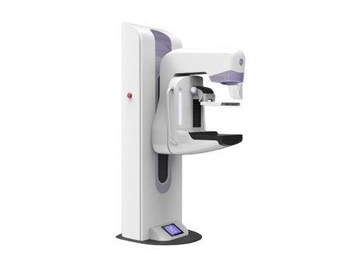
The Basic Principle of Digital Mammography
A Digital Mammography system uses low-dose X-rays to create detailed images of breast tissue. During an examination, the breast is positioned between two compression plates. Compression spreads the tissue evenly, reduces movement, and decreases the amount of radiation required to produce an image.
The X-ray tube generates a controlled beam that passes through the compressed breast. Different tissues absorb different amounts of X-ray energy, creating variations in the detected signal.
After passing through the breast, the remaining X-ray photons reach the digital detector. The detector converts these signals into electronic data, which is processed by specialized software to generate the final image.
Compared with conventional film-based mammography, digital imaging provides greater flexibility because image characteristics can be optimized after acquisition. Radiologists can adjust parameters such as brightness and contrast to better evaluate specific areas.
Key Components of a Digital Mammography System
A modern Digital Mammography system consists of several components that work together to achieve accurate breast imaging.
X-ray Generation System
The X-ray tube is responsible for producing the radiation required for imaging. Mammography systems use specially designed X-ray tubes to provide suitable energy levels for visualizing breast tissue.
Because breast abnormalities may appear as very small differences in tissue density, accurate control of X-ray energy is essential for maintaining image quality.
Compression System
Breast compression is an important part of mammography. It reduces tissue thickness and minimizes overlapping structures, allowing abnormalities to be detected more clearly.
Compression also helps reduce radiation exposure because less X-ray energy is required when the breast is evenly positioned.
Flat Panel Detector
The Flat Panel Detector (FPD) is one of the most important components in Digital Mammography. It captures the X-ray signals after they pass through breast tissue and converts them into digital information.
A high-quality detector must provide excellent sensitivity and spatial resolution because many breast abnormalities, such as microcalcifications, can be extremely small.
Detector performance affects several aspects of breast imaging, including:
- Fine detail visibility
- Contrast resolution
- Image consistency
- Radiation efficiency
The development of advanced Flat Panel Detector technology has significantly improved the performance of modern Mammography Digital Radiography systems.
Image Processing Software
After image acquisition, software processes the raw detector data and creates images suitable for clinical interpretation.
Modern image processing technologies help optimize contrast, reduce noise, and improve visualization of important structures. However, software performance depends on the quality of the original data captured by the detector.
The Role of Flat Panel Detector in Breast Imaging
The detector is a key factor that determines the performance of a Digital Mammography system.
Breast imaging presents unique challenges because many clinically important findings have very low contrast compared with surrounding tissue. A detector must capture small variations in X-ray absorption while maintaining low noise levels.
Modern Flat Panel Detector technology improves this process by providing efficient X-ray conversion and accurate signal collection.
For example, early-stage breast abnormalities may appear as tiny calcifications or small density changes. If the detector cannot capture these details clearly, important information may be lost during image acquisition.
This is why detector quality is one of the main considerations when evaluating a Mammography Digital Radiography system.
Conclusion
Digital Mammography has become an important technology in modern breast imaging by combining digital detector technology with advanced image processing. Its ability to capture subtle differences in breast tissue makes it valuable for both screening programs and diagnostic examinations.
When evaluating a mammography system, image quality, detector performance, workflow efficiency, and system adaptability are all important considerations. A well-designed system can help healthcare providers achieve reliable imaging performance while supporting long-term clinical needs.
At SEEHO Medical Equipment, we focus on developing and supplying medical imaging solutions that support different clinical environments. Our Mammography Digital Radiography systems are designed to provide stable image acquisition and practical workflow solutions for hospitals and diagnostic centers.

