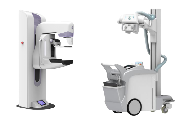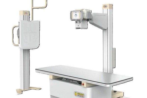
Software features in dynamic digital radiography typically include image acquisition controls, multi-speed cine playback, real-time image processing, and clinical data management. Specific tools vary by system model, software version, and vendor build. Modern software does much more than display static X-ray pictures on a screen. It controls pulsed exposures, suppresses image noise, and tracks motion changes over time. By turning rapid multi-frame sequences into clear diagnostic views, dynamic digital radiography software helps medical teams evaluate moving anatomy alongside traditional still images.
Dynamic Image Acquisition and Cine Playback
The acquisition console is the main workstation interface. Technologists use it to manage multi-frame cine runs during patient exams. The software sets exposure pulse rates and coordinates detector readouts so images stream smoothly without dropped frames.
After an acquisition, clinicians review the moving sequence with dedicated playback tools:
- Variable-Speed Review: Users can switch between normal speed, slow motion, and fast forward to inspect fast movements.
- Frame-by-Frame Stepping: Stepping through frames one by one lets doctors isolate the exact moment an articulation shifts.
- Continuous Cine Loops: Repeating forward-and-reverse loops makes it easy to track cyclic motions like breathing or joint flexion.
The software stores these dynamic sequences next to standard static views. This setup simplifies long-term archiving and helps clinicians compare follow-up studies side by side.
Image Processing and Enhancement
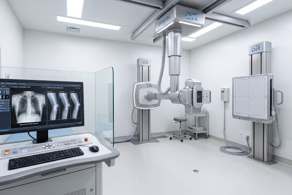
Raw dynamic projection data often contains electronic noise, scatter radiation, and low contrast. Dynamic digital radiography software uses fast processing algorithms to clean up the image without blurring moving edges.
Noise filters suppress frame-to-frame grain, while edge enhancement tools sharpen bone borders and lung lines. Dynamic range adjustments balance bright bones and darker soft tissues so both remain clearly visible. However, software filters cannot fix bad raw data. If a dynamic digital radiography exam is underexposed or blurred by poor positioning, software sharpening will only make the background noise worse.
| Software Module | Primary Function | Core Clinical Benefit |
| Acquisition & Playback | Sets pulsed rates and provides review controls | Enables smooth motion viewing and frame-by-frame inspection |
| Image Processing | Applies noise filters and contrast balancing | Removes grain while keeping bone edges and soft tissue sharp |
| Motion Analysis | Tracks anatomical landmarks across frames | Generates objective displacement curves and movement charts |
| Data Management | Handles DICOM transfer and local storage | Simplifies archiving, report sharing, and follow-up comparisons |
Motion Analysis and Quantitative Measurement Tools
Advanced dynamic digital radiography systems include automated tools that extract objective measurements from moving image loops. These features turn visual movement into clear numerical data.
Technologists can place a designated marker or select a region of interest around specific anatomy. The software then tracks how those pixels move across consecutive frames, plotting displacement curves, velocity data, and range-of-motion charts. In chest studies, for example, the software traces the vertical movement of the diaphragm during deep breathing. These quantitative outputs provide objective reference points, though clinicians must still verify that patient position remained steady throughout the exam.
Transforming Dynamic Data into Clinical Information
Software tools in dynamic digital radiography turn raw image frames into useful clinical information by organizing motion data into straightforward diagnostic views.
A single dynamic sequence generates hundreds of separate frames. Reviewing them one by one without good software would take too much time. Playback controls let doctors scan an entire motion cycle in seconds, while analytical tools highlight subtle motion problems that are easy to miss. Even so, software measurements should never replace clinical judgment. Automated curves simply assist the radiologist, who must interpret the findings against the patient’s symptoms and medical history. Because software packages range from basic viewers to advanced measurement suites, buyers should test workstation features carefully before choosing a platform.
Conclusion
Software in dynamic digital radiography covers image acquisition, cine playback, noise filtering, motion measurement, and data storage. It serves as an essential bridge, turning rapid X-ray frames into practical information about anatomical motion and structure. Because software tools differ between manufacturers, medical facilities should review specific workstation capabilities to match their clinical workflow and diagnostic needs.
Contact our technical imaging team today to schedule an interactive software demonstration and explore dynamic digital radiography solutions configured for your facility.