
Can Dynamic Digital Radiography Capture Real-Time Motion?
Dynamic digital radiography captures real-time physiological motion across multiple organ systems. The system records sequential diagnostic frames under active, weight-bearing conditions without inducing motion smear.
Conventional projection radiography produces sharp anatomical snapshots. However, static radiographs depict human anatomy in completely frozen states. Many musculoskeletal and pulmonary conditions cause symptoms exclusively during physical movement. Clinicians often miss dynamic joint impingement, occult subluxation, and asymmetric diaphragm excursion on standard views.
Fluoroscopy provides real-time guidance during interventions, yet it generates lower diagnostic resolution alongside noticeable image lag. To overcome these diagnostic obstacles, modern imaging facilities utilize dynamic digital radiography. This advanced modality combines the fine spatial detail of digital radiography with continuous motion acquisition. Clinicians can observe living biomechanics while keeping cumulative radiation doses comparable to standard examinations.
High-Speed Acquisition Versus Fluoroscopy in Dynamic Digital Radiography
The motion-capture capabilities of dynamic digital radiography depend directly on high-speed flat-panel detector architecture. Conventional fluoroscopy systems operate primarily as procedural guidance tools. Consequently, fluoroscopy sacrifices sharp spatial resolution to maintain video frame rates.
Fluoroscopic detectors also suffer from capacitive image lag. Moving bones leave faint trails or ghost margins across successive display frames. In contrast, dynamic digital radiography utilizes ultra-short exposure pulses paired with rapid thin-film transistor readout arrays.
| Imaging Parameter | Conventional Fluoroscopy | Dynamic Digital Radiography (DDR) |
| Frame Acquisition Rate | Standard video rates | 15 to 30 frames per second |
| Spatial Resolution | Low to moderate visual guidance | High-resolution diagnostic radiography |
| Pulse Duration per Frame | Continuous beam or long pulses | 1 to 5 ms ultra-short exposure bursts |
| Motion Smear & Lag | Noticeable trailing ghost artifacts | Complete elimination of image lag |
| Patient Posture | Typically recumbent positioning | Upright, physiological weight-bearing |
Modern pulsed radiographic platforms eliminate motion blur completely:
- A high-frequency X-ray generator discharges radiation pulses lasting only a few milliseconds per frame.
- The ultra-short pulse duration freezes fast anatomical displacement without geometric edge blurring.
- Fast cesium iodide scintillators convert incoming photons into visible light waves almost instantaneously.
- Specialized readout electronics clear residual charges between exposures, preventing trailing motion artifacts.
By synchronizing short radiation bursts with detector refresh cycles, functional radiographic imaging captures rapid biomechanical transitions clearly.
Musculoskeletal Biomechanical Tracking Using Motion-Tracking Radiography
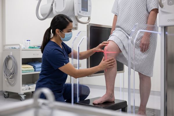
Evaluating joint performance under physiological stress remains a core strength of dynamic digital radiography. Traditional static X-rays force patients into resting, non-weight-bearing postures. These unnatural configurations mask mechanical instabilities that emerge solely during active weight-bearing maneuvers.
Kinetic radiographic imaging records joints through complete physical flexion and extension cycles:
- Orthopedic specialists evaluate cervical spine instability by tracking vertebrae throughout active flexion and extension.
- Radiologists assess patellofemoral tracking abnormalities as the patient performs natural, weight-bearing squats.
- Clinicians detect dynamic shoulder impingement during active arm abduction, pinpointing painful subacromial friction zones.
- Technologists record real-time wrist motions, identifying scapholunate dissociation and subtle ligamentous laxity immediately.
Surgeons also employ functional digital X-rays to assess complex post-surgical outcomes:
- The system tracks prosthetic joint kinetics, revealing subtle implant loosening during active ambulatory loading.
- Radiologists identify micro-motion along bone-implant interfaces long before structural osteolysis appears on static films.
- Clinicians differentiate soft tissue adhesions from true hardware failure during symptomatic motion arcs.
- Physicians design targeted physical therapy regimens based on objective joint displacement measurements.
By recording joints under true weight-bearing loads, dynamic digital radiography exposes hidden mechanical dysfunction reliably.
Real-Time Thoracic and Pulmonary Dynamics in Functional Radiographic Imaging
Thoracic disorders frequently distort pulmonary ventilation mechanics without creating dense structural lesions. Static chest radiographs show gross parenchymal consolidation, but they cannot evaluate active respiratory mechanics. Clinicians rely on dynamic digital radiography to visualize breathing dynamics across complete ventilatory cycles.
Pulsed dynamic imaging tracks the moving chest wall and lungs throughout forced inspiration and expiration:
- Radiologists trace continuous bilateral diaphragmatic excursion, quantifying vertical craniocaudal displacement in real time.
- The system measures diaphragmatic velocities, confirming phrenic nerve palsy when paradoxical elevation occurs during inhalation.
- Clinicians evaluate dynamic tracheal collapse, diagnosing tracheobronchomalacia as airway diameters shrink during exhalation.
- Physicians calculate regional lung area changes, identifying localized hypoventilation across individual pulmonary lobes.
This functional data proves exceptionally valuable for chronic obstructive pulmonary disease (COPD) management:
- Dynamic X-ray sequences visualize air-trapping kinetics, demonstrating delayed pulmonary deflation during forced expiration.
- Radiologists detect abnormal chest wall excursion, identifying intercostal muscle recruitment and paradoxical rib cage motion.
- Pulmonologists monitor therapeutic responses by comparing diaphragmatic movement metrics before and after bronchodilator administration.
- Clinicians gain actionable functional insights without subjecting frail patients to complex plethysmography chambers.
Consequently, dynamic digital radiography transforms routine thoracic examinations into quantitative assessments of respiratory biomechanics.
Advance Clinical Care With Functional Motion-Tracking Systems
Conventional static imaging forces physicians to infer dynamic dysfunction from motionless anatomical projections. In contrast, dynamic digital radiography provides direct kinetic confirmation, preventing diagnostic delays and unnecessary exploratory procedures. Medical facilities equip their departments with functional motion radiography to resolve complex orthopedic complaints and subtle pulmonary disorders decisively.
Are you ready to expand your imaging capabilities beyond static boundaries? Our innovative dynamic digital radiography systems empower radiologists with objective kinetic data, shorter patient turnaround times, and superior diagnostic clarity. Reach out to our application specialists today to explore clinical workflow integration, view kinetic imaging datasets, and secure your departmental consultation.

