
Dynamic Digital Radiography for Orthopedic Assessment
Dynamic Digital Radiography for orthopedic assessment is gaining increasing attention as clinicians seek imaging techniques that provide both anatomical and functional information. Conventional Digital Radiography remains the first-line imaging method for evaluating fractures, bone alignment, and joint structure. However, many orthopedic conditions are associated with abnormal motion rather than obvious structural changes, meaning patients may experience pain, instability, or limited mobility even when static radiographs appear normal.
Dynamic Digital Radiography (DDR) addresses this challenge by capturing a continuous sequence of digital X-ray images during movement. Rather than recording a single moment, it visualizes how bones and joints function throughout an activity, providing additional information that complements conventional radiographic findings.
Why Dynamic Digital Radiography Matters in Orthopedic Assessment
Many musculoskeletal disorders develop during movement. Joint instability, abnormal kinematics, and altered biomechanics may not be visible when the patient remains still for a conventional X-ray examination.
Dynamic Digital Radiography addresses this limitation by allowing joints to be evaluated while they move through a controlled range of motion. The examination produces a continuous image sequence, making it possible to observe joint mechanics as they occur rather than inferring movement from static images.
This additional functional information can improve clinical understanding, particularly when conventional radiographs fail to explain a patient’s symptoms.
Dynamic Digital Radiography for Joint Motion Analysis
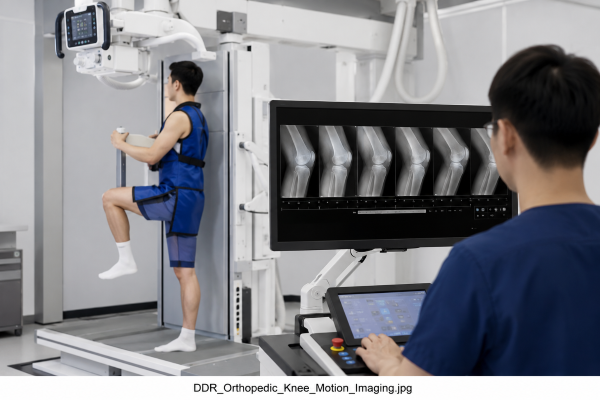
Evaluating joint motion is one of the most promising orthopedic applications of DDR. Recent studies have investigated its use in assessing shoulder movement, cervical spine mobility, gait analysis, and postoperative joint kinematics.
For example, researchers have used Dynamic Digital Radiography to measure shoulder motion throughout arm elevation. Instead of relying solely on visual interpretation, the examination allowed quantitative analysis of scapular and humeral movement during abduction. These measurements provided objective data that supported functional assessment of the shoulder.
Because the examination captures the entire movement process, clinicians can evaluate motion patterns that are impossible to observe on a single radiograph.
Supporting Postoperative Orthopedic Evaluation
Successful orthopedic surgery is measured not only by implant position but also by functional recovery. Although conventional Digital Radiography remains essential for postoperative imaging, static images cannot demonstrate how an operated joint performs during movement.
Dynamic Digital Radiography has therefore been investigated as a complementary imaging technique after orthopedic procedures. Published studies have reported its use in evaluating joint kinematics following total hip arthroplasty and total knee arthroplasty. Dynamic image sequences helped identify mechanical abnormalities that were difficult to appreciate using conventional radiographs alone, providing additional information for postoperative assessment.
Rather than replacing standard X-ray examinations, DDR adds another layer of clinical information that may support treatment planning and rehabilitation.
Potential Applications in Sports Medicine
Sports injuries often involve repetitive movement, joint instability, or altered biomechanics. These functional abnormalities may only become apparent while an athlete is performing a specific motion.
Dynamic Digital Radiography provides an opportunity to evaluate movement under controlled conditions. Researchers have explored its application in shoulder biomechanics and other musculoskeletal assessments where quantitative motion analysis is clinically valuable. By visualizing movement frame by frame, clinicians can better understand how a joint functions throughout an activity instead of evaluating only its resting position.
As research continues, DDR may become an increasingly useful tool for monitoring rehabilitation progress and guiding return-to-play decisions.
The Role of DDR in Modern Orthopedic Imaging
Dynamic Digital Radiography is not intended to replace conventional Digital Radiography, CT, or MRI. Each imaging modality serves a different clinical purpose.
Static radiographs remain indispensable for evaluating bone structure, while MRI continues to provide detailed soft tissue assessment. DDR fills a different role by adding functional visualization to routine X-ray imaging. This combination of anatomical and motion information allows clinicians to examine both how a joint looks and how it performs during movement.
As evidence continues to grow, Dynamic Digital Radiography is becoming an important complement to existing orthopedic imaging techniques rather than an alternative to them.
Conclusion
Orthopedic diagnosis often requires more than identifying structural abnormalities. Understanding how joints move can provide valuable information that is not available from conventional radiographs alone.
Dynamic Digital Radiography expands the capabilities of Digital Radiography by introducing real-time functional assessment into routine X-ray imaging. Current research has demonstrated promising applications in joint motion analysis, postoperative evaluation, shoulder biomechanics, and gait assessment. As clinical experience and imaging technology continue to advance, DDR is expected to play an increasingly important role in orthopedic assessment.
At SEEHO Medical Equipment, we are committed to providing advanced Digital Radiography solutions that support accurate diagnosis and efficient clinical workflows. If you would like to learn more about our Dynamic Digital Radiography systems or discuss your imaging requirements, our team is ready to provide professional advice and tailored solutions.

