
What Is a Flat Panel Detector and How Does It Work?
A Flat Panel Detector (FPD) is a digital imaging device that converts X-rays into electronic signals to produce digital medical images. As the core imaging component of modern Digital Radiography Systems, it captures X-rays after they pass through the patient’s body and transfers the imaging data to the system’s software for image reconstruction. This digital process enables healthcare professionals to obtain high-quality X-ray images within seconds, improving diagnostic efficiency, image quality, and clinical workflow.
As digital radiography has become the standard in modern medical imaging, Flat Panel Detectors have largely replaced traditional X-ray film and Computed Radiography (CR) technology. Understanding how a Flat Panel Detector works helps healthcare providers, distributors, and equipment buyers better understand the technology behind Digital Radiography Systems and make more informed equipment decisions.
The Working Principle of a Flat Panel Detector
Although a digital X-ray image appears on the workstation within seconds, several processes take place before the image is generated. From X-ray exposure to digital image reconstruction, each step plays an important role in producing accurate diagnostic images.
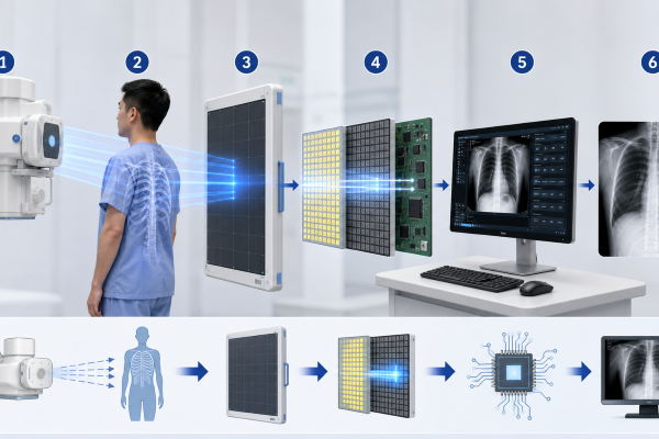
X-ray Generation
The imaging process begins when the X-ray tube generates X-rays using a controlled amount of radiation based on the selected examination parameters. These X-rays travel toward the patient and carry the information required to create a diagnostic image.
X-ray Transmission Through the Patient
As the X-rays pass through the patient’s body, different tissues absorb different amounts of radiation according to their density. Dense structures such as bones absorb more X-rays, while soft tissues allow more X-rays to pass through.
These differences in X-ray absorption create the anatomical information that forms the basis of the final medical image.
X-ray Detection
After passing through the patient, the remaining X-rays reach the Flat Panel Detector.
The detector captures the transmitted X-ray energy across its imaging surface while preserving the information carried by the X-rays. At this stage, the detector has collected the raw imaging data, but the information has not yet been converted into a visible digital image.
Signal Conversion
The captured X-ray energy is then converted into electronic signals inside the Flat Panel Detector.
Depending on the detector technology, this conversion may occur through either a direct or indirect process. Regardless of the method used, the objective is to transform X-ray energy into digital data that can be processed by the imaging system.
Digital Image Processing
The electronic signals are transmitted to the Digital Radiography System, where advanced image processing software reconstructs the final image.
During this process, the software automatically performs multiple optimization tasks, including noise reduction, contrast enhancement, edge sharpening, and image correction. These processing steps help improve image clarity and support accurate clinical diagnosis.
Image Display and Storage
Once image processing is complete, the digital X-ray image is displayed on the workstation within a few seconds.
The image can then be reviewed immediately by clinicians, stored in the hospital’s PACS, or shared electronically with other healthcare professionals, enabling a faster and more efficient diagnostic workflow.
Types of Flat Panel Detector Technology
Although all Flat Panel Detectors perform the same fundamental task of converting X-rays into digital images, they use different technologies to achieve signal conversion. Modern Flat Panel Detectors are generally classified into two main types.
Indirect Conversion Technology
Indirect conversion technology first converts X-rays into visible light using a scintillator material, most commonly cesium iodide (CsI) or gadolinium oxysulfide (GOS). The visible light is then converted into electronic signals by a photodiode layer, typically made of amorphous silicon (a-Si).
This technology is widely used in general Digital Radiography because it provides excellent image quality, stable performance, and high dose efficiency.
Direct Conversion Technology
Direct conversion technology uses amorphous selenium (a-Se) to convert X-ray energy directly into electronic signals without first producing visible light.
Because it eliminates the light conversion stage, direct conversion technology minimizes light spread and can achieve higher spatial resolution, making it particularly suitable for applications that require exceptional image detail.
Both technologies are widely used in modern Digital Radiography Systems, and the most appropriate choice depends on clinical applications, imaging requirements, and system design.
Key Components of a Flat Panel Detector
A Flat Panel Detector consists of several key components that work together to capture X-rays and generate digital images. Each component performs a specific function, ensuring accurate signal conversion and reliable imaging performance.
Protective Layer
The protective layer is the outer surface of the detector. It shields the internal components from physical impact, dust, and daily wear while allowing X-rays to pass through with minimal attenuation. A durable protective layer helps extend the service life of the detector in busy clinical environments.
Scintillator or Conversion Layer
The conversion layer is responsible for converting X-ray energy into a form that can be processed by the detector.
- In indirect conversion detectors, the scintillator (such as cesium iodide or gadolinium oxysulfide) converts X-rays into visible light.
- In direct conversion detectors, an amorphous selenium (a-Se) layer converts X-rays directly into electronic charges.
This layer plays a critical role in determining image quality and detector performance.
Photodiode Layer
In indirect conversion detectors, the photodiode layer receives the visible light produced by the scintillator and converts it into electronic signals. Amorphous silicon (a-Si) is commonly used because of its stability and reliable imaging performance.
Thin Film Transistor (TFT) Array
The Thin Film Transistor (TFT) array collects the electronic signals generated across the detector surface and transfers them to the readout electronics.
Each pixel has its own transistor, allowing the detector to accurately record image information with high resolution and fast readout speed.
Readout Electronics
The readout electronics receive the signals collected by the TFT array and convert them into digital data that can be processed by the imaging software.
This process enables the Digital Radiography System to reconstruct the final medical image for clinical diagnosis.
Why Flat Panel Detector Technology Matters
Flat Panel Detector technology is fundamental to modern Digital Radiography because it directly influences how efficiently X-ray information is captured and converted into diagnostic images.
A high-performance detector contributes to:
- High-quality digital images for accurate diagnosis
- Fast image acquisition and shorter examination times
- Efficient digital workflow within healthcare facilities
- Stable and reliable imaging performance
- Improved overall efficiency of Digital Radiography Systems
For these reasons, the Flat Panel Detector is widely regarded as the core imaging component of modern Digital Radiography Systems.
Conclusion
A Flat Panel Detector is the key component that enables modern Digital Radiography Systems to produce high-quality digital X-ray images. By capturing X-rays, converting them into electronic signals, and supporting advanced image processing, it plays a vital role in diagnostic accuracy and clinical efficiency.
Understanding the working principle, detector types, and internal components of a Flat Panel Detector provides a solid foundation for understanding how Digital Radiography technology functions in today’s healthcare environment.
If you are planning to invest in a Digital Radiography System equipped with advanced Flat Panel Detector technology, welcome tocontact us. Our professional team can recommend suitable imaging solutions and provide technical support based on your clinical requirements and project goals.

